
Research Article 
 Creative Commons, CC-BY
Histological Analysis of the Cardiac Fulcrum and the Atrio Ventricular Node of the Heart and its Functional Implication
*Corresponding author:Jorge Carlos Trainini, Residente Perón Hospital. National University of of Avellaneda, Argentina
Received:October 16, 2026; Published:October 23, 2026
DOI: 10.34297/AJBSR.2026.32.004191
Abstract
Objective: We have studied the relationship of the Cardiac Fulcrum in its anatomical location, without unfolding the heart into its muscular band arrangement. The objective is to observe the relationships of the Fulcrum with the other anatomical elements.
Material and Methods: The methods used in this research to explain the hypothesis of the anatomy functional integrity of the heart of: 1. The use of consisted 84 hearts sourced from morgues, slaughterhouses (bovine, humans and porcine). 2. Histological and histochemical analysis of the anatomical samples.
Results: We observed that in both bovine and porcine models, as well as in humans, the Fulcrum presents a configuration featuring a central area and two poles. A pointed right pole inserts into the velamentous sector of the tricuspid valve, and a left pole-ovoid in bovines or forming a double arch in porcine- inserts into the ascending loop of the left ventricle and is in intimate contact with the atrio ventricular node. The central zone of the Fulcrum constitutes the lever arm. This configuration allows the Fulcrum to transmit a pulsatile rocking motion to the atrio ventricular node. The Fulcrum and the atrio ventricular node regulate and modulate the afferent information from the organism before converting it into the efferent depolarization wave. Therefore, the Fulcrum is the physical component that allows the electrical system or depolarization waves to couple or decouple (modulate), providing the myocardium with an efferent message in response to the afferent messages received from all somatic nodes that report on the internal and external environment of the organism.
Conclusions: The final conclusion is that the fulcrum has a dual function. First, it supports the double helix that shapes the anatomy of the heart; and second, it acts as a transmitter by integrating with the atrioventricular node and the autonomic nervous system.
Keywords:Cardiac fulcrum, Node AV, Helical heart
Introduction
In a series of congresses and meetings held throughout the 1960s, Francisco Torrent Guasp proposed the concept that the heart consists of a single muscular band folded upon itself to form a double helix [1-3]. Despite numerous meetings and presentations, it proved impossible to shift the prevailing paradigm, which viewed the heart as an organ with a solid myocardium. Perhaps the primary criticism centered on the absence of an anchor point from which contractile movements could originate. Years later, Jorge Trainini identified such a point in both bovine and human hearts [4-6]; this research was published in the journal
*Morphology* in 2021 [5]. This structure, termed a *fulcrum* (a point of support), is composed of osteochondroid tissue in cattle, and of cartilaginous tissue in pigs and humans. However, unrolling the cardiac muscular band alters all its anatomical relationships-including those it maintains with the *fulcrum* and the atrioventricular node, the site where the muscular band inserts into the *fulcrum*. Furthermore, the atrioventricular node has not been the subject of sufficient histological study regarding its relationship to the *fulcrum*. The Purkinje cells, discovered in 1830, were also investigated; these cells are thought to conduct energy, thereby accelerating the propagation of the energetic wave [7-12]. Consequently, these relationships were analyzed-relationships which, according to our observations, have not yet been sufficiently documented in the scientific literature. This article also evaluates the physiological implications of these findings.
Materials and Methods
The methods used in this research to explain the hypothesis of
the anatomy functional integrity of the heart of:
A. The use of consisted 84 hearts sourced from morgues,
slaughterhouses:
a) 57 two-year-old bovine hearts weighing between 1300
and 1900 g (average 1650 g);
b) 17 human hearts (three at 8, 16, and 23 weeks of gestation,
respectively; four infants at 30 days, 36 days, 10 weeks, and 27
weeks of age; one 4-year-old child; one 10-year-old child; and
eight adults with an average weight of 300 g);
c) 10 porcine hearts (400 g);
B. Histological and histochemical analysis of the anatomical
samples. The study will be performed using hematoxylin and
eosin staining and Masson’s trichrome staining. Immunostaining
techniques for neurofilaments will also be performed.
Results
The results are presented in the histological figures of the research conducted (Figures 1-8).
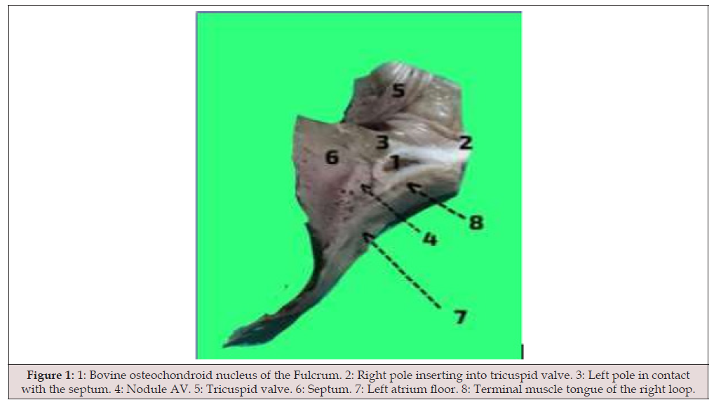
Figure 1:1: Bovine osteochondroid nucleus of the Fulcrum. 2: Right pole inserting into tricuspid valve. 3: Left pole in contact with the septum. 4: Nodule AV. 5: Tricuspid valve. 6: Septum. 7: Left atrium floor. 8: Terminal muscle tongue of the right loop.
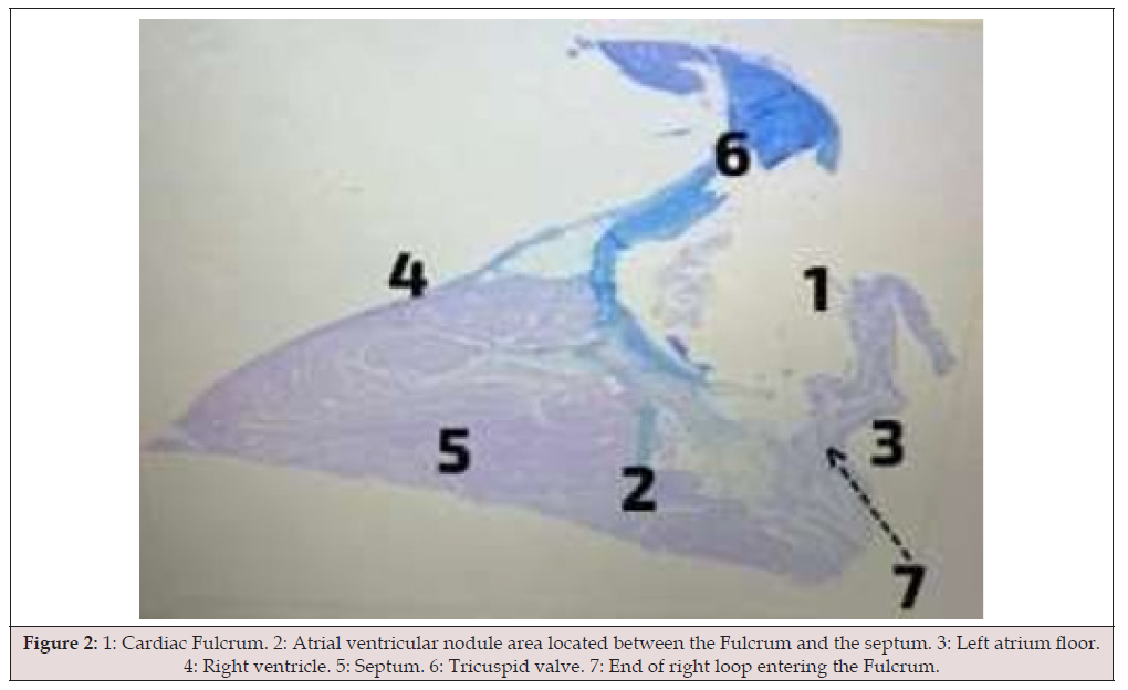
Figure 2:1: Cardiac Fulcrum. 2: Atrial ventricular nodule area located between the Fulcrum and the septum. 3: Left atrium floor. 4: Right ventricle. 5: Septum. 6: Tricuspid valve. 7: End of right loop entering the Fulcrum.
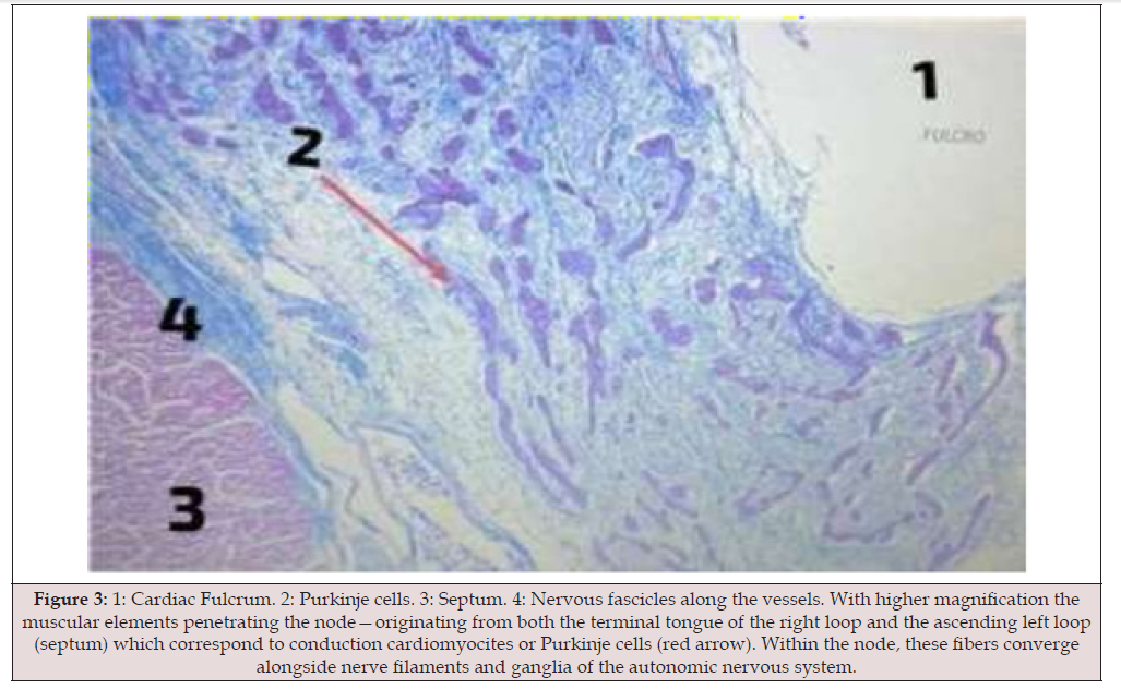
Figure 3:1: Cardiac Fulcrum. 2: Purkinje cells. 3: Septum. 4: Nervous fascicles along the vessels. With higher magnification the muscular elements penetrating the node—originating from both the terminal tongue of the right loop and the ascending left loop (septum) which correspond to conduction cardiomyocites or Purkinje cells (red arrow). Within the node, these fibers converge alongside nerve filaments and ganglia of the autonomic nervous system.
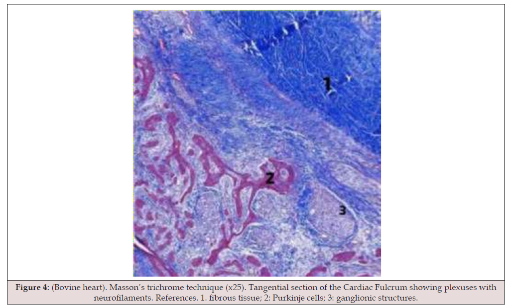
Figure 4:(Bovine heart). Masson’s trichrome technique (x25). Tangential section of the Cardiac Fulcrum showing plexuses with neurofilaments. References. 1. fibrous tissue; 2: Purkinje cells; 3: ganglionic structures.
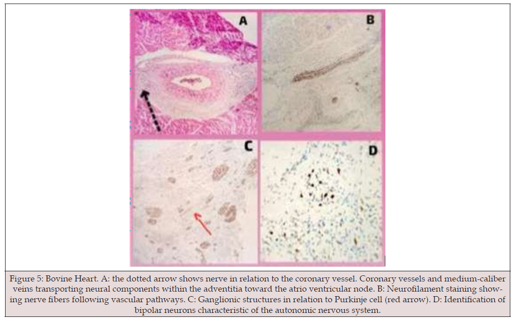
Figure 5:Bovine Heart. A: the dotted arrow shows nerve in relation to the coronary vessel. Coronary vessels and medium-caliber veins transporting neural components within the adventitia toward the atrio ventricular node. B: Neurofilament staining showing nerve fibers following vascular pathways. C: Ganglionic structures in relation to Purkinje cell (red arrow). D: Identification of bipolar neurons characteristic of the autonomic nervous system.
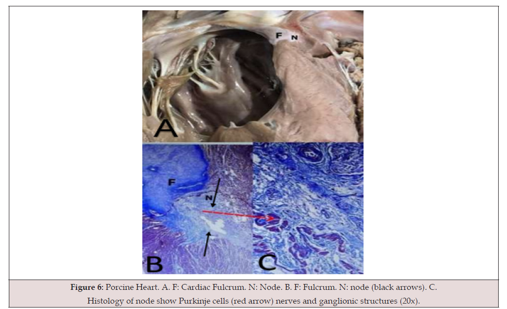
Figure 6:Porcine Heart. A. F: Cardiac Fulcrum. N: Node. B. F: Fulcrum. N: node (black arrows). C. Histology of node show Purkinje cells (red arrow) nerves and ganglionic structures (20x).
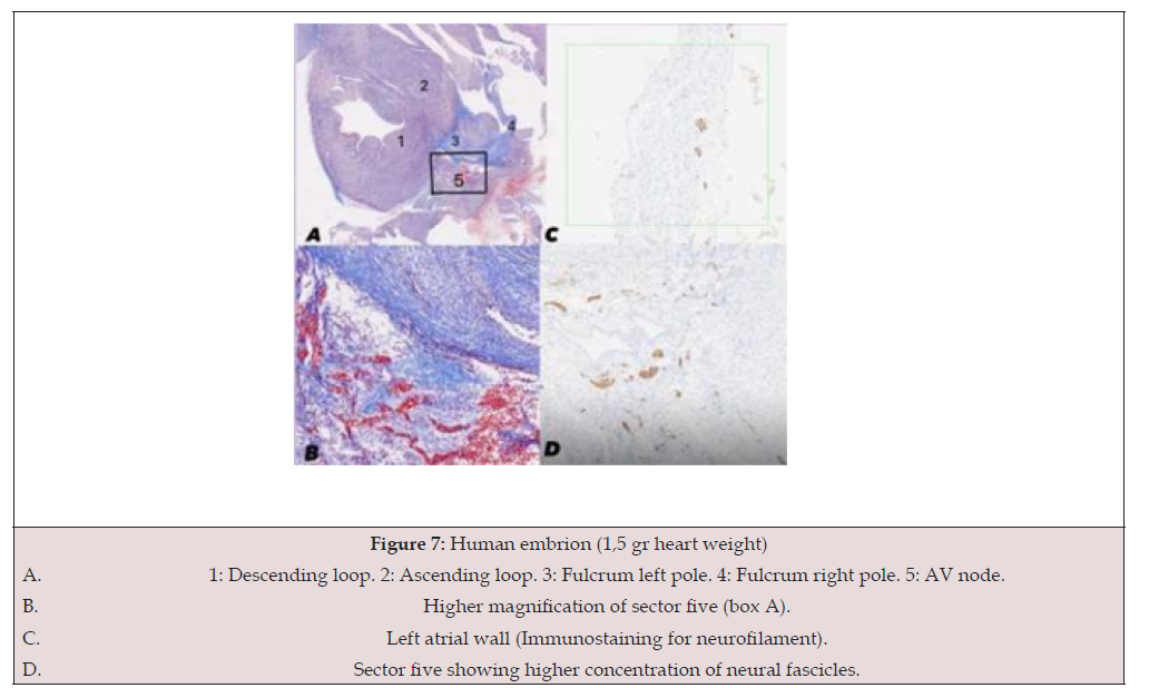
Figure 7:Human embrion (1,5 gr heart weight)
A. 1: Descending loop. 2: Ascending loop. 3: Fulcrum left pole. 4: Fulcrum right pole. 5: AV node.
B. Higher magnification of sector five (box A).
C. Left atrial wall (Immunostaining for neurofilament).
D. Sector five showing higher concentration of neural fascicles.
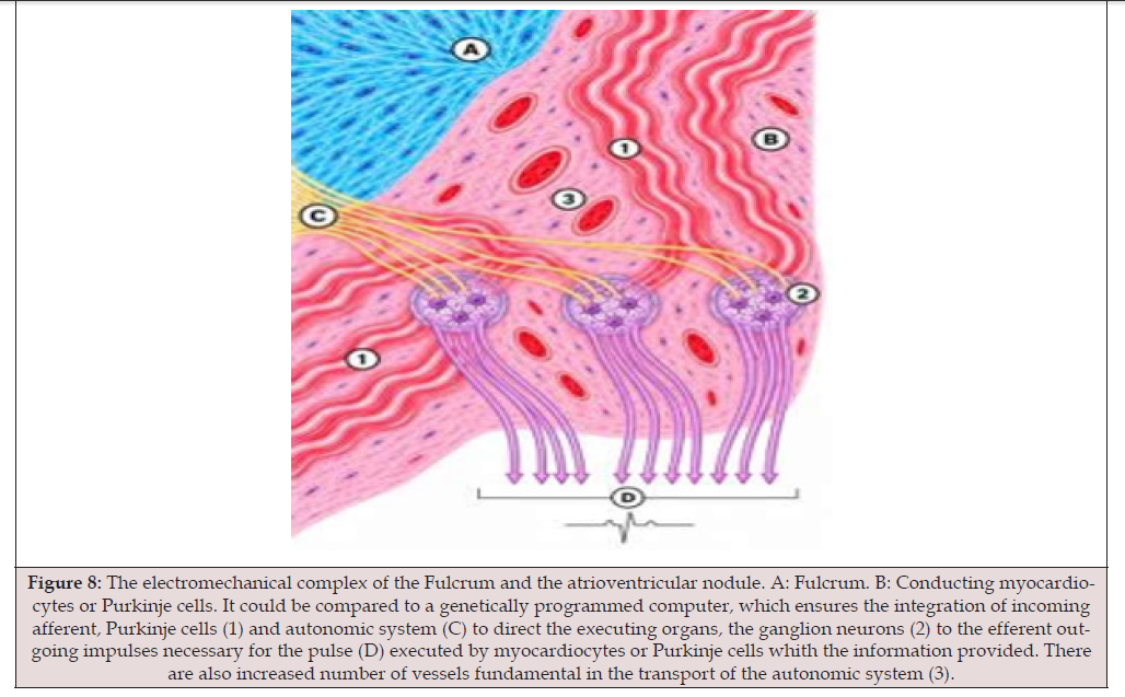
Figure 8:The electromechanical complex of the Fulcrum and the atrioventricular nodule. A: Fulcrum. B: Conducting myocardiocytes or Purkinje cells. It could be compared to a genetically programmed computer, which ensures the integration of incoming afferent, Purkinje cells (1) and autonomic system (C) to direct the executing organs, the ganglion neurons (2) to the efferent outgoing impulses necessary for the pulse (D) executed by myocardiocytes or Purkinje cells whith the information provided. There are also increased number of vessels fundamental in the transport of the autonomic system (3).
Discussion
In this study, we investigated the fulcrum and its relationship to cardiac innervation, while preserving all anatomical relationships. These relationships are altered during the unfolding of the heart-a process essential for understanding that the heart is composed of a single muscular band arranged as a double helix. Furthermore, the study of the fulcrum revealed a rich innervation by the autonomic nervous system. Bovine, porcine, and human (both adult and embryonic) hearts were examined. In general, the fulcrum consists of two poles-left and right-and an arm approximately 3 cm in length (varying by species), with specific interrelationships among these components. The right pole is pointed, whereas the left pole is oval-shaped in bovine hearts, or forms an irregular double arch at its terminal end in porcine and human hearts. This configuration arises from the specific relationships between the two poles: the right pole inserts into the velamentous portion of the tricuspid valve, while its counterpart, the left pole, is in close contact with the insertion of the ascending limb of the left ventricle. In bovine hearts, the fulcrum features a central zone composed of osteochondroid tissue, which appears as a dark brown, teardrop-shaped area in (Figure 1).
The midsection of the bovine fulcrum runs along the floor of the left atrium. Between the fulcrum and the floor of the left atrium lies a muscular band-corresponding to the terminal muscular fascicle of the right loop-which will be discussed further on. We have observed a dense network of nerves that follows the course of the blood vessels. This innervation belongs to the autonomic nervous system and reaches the heart via the adventitia of the superior and inferior venae cavae and the coronary vessels, arriving at the Atrioventricular (AV) node through a corridor of loose connective tissue that covers the surface adjacent to the ventricular septum and the AV node. Thus, the autonomic nervous system-by way of the superior vena cava-incorporates stimuli originating from the brain, while, via the inferior vena cava, it incorporates stimuli from the gastrointestinal and urogenital systems. Consequently, there exists a somatic connection, mediated by the autonomic nervous system, which is centered on the heart.
In both bovine and porcine hearts, we have observed that this innervation follows the path of the blood vessels and terminates at the atrioventricular node. This node is influenced by pulsatile movement; in this way, a pulsatile oscillation is transmitted through this “pivot point” (or fulcrum), which can regulate and modulate the afferent and efferent somatic stimuli acting upon the atrioventricular node. Afferent stimuli originate throughout the somatic
environment, while efferent stimuli are those that generate the wave of depolarization. This function constitutes a regulatory modulation-a temporal regulation of the electromechanical information that this pivot point exerts upon the atrioventricular node. Therefore, this central node, located at the core of the heart, is connected to other nodes within the complex somatic network that constitutes a living being.
It is now appropriate to examine the histology of the atrioventricular node. The histology of the AV node reveals a cell type derived from two distinct lineages of cardiomyocytes: specifically, those originating from the terminal muscular fascicle of the right branch and those derived from the left ascending branch. These conducting cardiomyocytes, also known as Purkinje cells, are wider, exhibit less pronounced transverse striation, and display a distinct scalloped pattern. These cells are arranged within a network of autonomic ganglia and nerve tracts that make up the bulk of the node. The distinctive staining properties of these cells, combined with their marked scalloped pattern, suggest that the peripheral ganglionic environment influences their morphology.
Finally, in a cross-section of the embryonic heart, it can be inferred that the contraction of the left ventricle-with its two layers-functions analogously to a ball bearing. These movements provide continuous lubrication to the myocardium throughout cycles of contraction and relaxation, forcing a hyaluronic acid-rich plasma into the spaces between the cardiomyocytes [10]. Using staining techniques on the embryonic heart, we observed multiple ganglionic elements that traverse the wall of the right atrium and reach the cardiac base-located beneath the floor of the left atriumindicating the presence of components of the autonomic nervous system. In other words, they are found at the cardiac base, below the floor of the left atrium.
The fulcrum and the AV node regulate and modulate stimuli arriving from the entire body (soma) before converting them into the depolarization wave. It combines afferent information-what the body senses (cerebral, gastric, urogenital stimuli)-that reaches the ganglion and Purkinje cells of the AV node and generates an efferent response, which is the electrical command to contract. The cardiac fulcrum can be conceptualized as an electromechanical coupling center where the helical geometry of the myocardium modulates and expresses electrical propagation and mechanical dynamics, generating a spatially coherent electromagnetic information field. Therefore, the cardiac fulcrum and the AV node constitute a transition point where electrical energy is converted into organized biological time.
Conclusions
This work studies the fulcrum and its anatomical relationships. The final conclusion is that the fulcrum has a dual function. First, it supports the double helix that shapes the anatomy of the heart; and second, it acts as a transmitter by integrating with the atrioventricular node and the autonomic nervous system. Each heartbeat is, therefore, the result of interpreting fluctuations from an external and internal environment, which enter the atrioventricular node-which acts almost as a second brain at the base of the heart-via the autonomic nervous system, in synchrony with the fulcrum. This generates rhythmic feedback with coupling and decoupling. The cardiac fulcrum can be conceptualized as an electromechanical coupling center where the helical geometry of the myocardium modulates and expresses electrical propagation and mechanical dynamics, generating a spatially coherent electromagnetic information field.
Acknowledgements
None.
Conflict of Interest
None.
References
- Torrent Guasp F (1998) Estructura y función del corazó Rev Esp Cardiol 51: 91-102.
- Torrent Guasp F, Buckberg G, Carmine C, Cox J, Coghlan H, Gharib M (2001) The structure y function of the helical heart y its buttress wrapping. I. The normal macroscopic structure of the heart. Seminars in Thorac y Cardiovasc Surg 13(4): 301-319.
- Trainini JC, Lowenstein JA, Beraudo M, Mora Llabata V, Carreras Costa F, et al. (2022) Fulcrum y Torsion of the Helical Myocardium. Biblos Ed, Buenos Aires.
- Trainini JC, Lowenstein J, Beraudo M, Valle Cabezas J, Wernicke M, et al. (2023) “Anatomy y organization of the helical heart”. UNDAV Ed, Buenos Aires.
- Trainini JC, Lowenstein J, Beraudo M, Wernicke M, Trainini A, et al. (2021) Myocardial torsion y cardiac fulcrum (Torsion myocardique et pivot cardiaque). Morphologie 105(348): 15-23.
- Arvidsson M, Töger J, Carlsson M, Steding Ehrenborg K, Pedrizzetti G, et al. (2017) Left and right ventricular hemodynamic forces in healthy volunteers and elite athletes assessed with 4D flow magnetic resonance imaging. Am J Physiol Heart Circ Physiol 312(2): H314-H328.
- Trainini Jorge, Beraudo Mario, Wernicke Mario, Trainini Alejandro, Lowenstein Jorge, et al. (2024) Fundamental Conclusions on Research into the Anatomy and Organization of the Helical Heart. Am J Biomed Sci Res 23(5): 647-657.
- Carreras F, Ballester M, Pujadas S, Leta R, Pons Lladó G (2006) Morphological and functional evidences of the helical heart from non-invasive cardiac imaging. Eur J Cardiothoracic Surg 29 (Suppl 1): S50-S55.
- Trainini JC, Beraudo M, Wernicke M, Carreras Costa F, Trainini A, et al. (2022) Evidence that the myocardium is a continuous helical muscle with one insertion. REC: CardioClinics 57: 194-202.
- Trainini JC, Wernicke M, Beraudo M, Lowenstein J, Trainini A, et al. (2021) Hyaluronic acid in the heart. J Trends Biomedical Research 4(1).
- Trainini JC, Beraudo M, Wernicke M, Carreras Costa F, Trainini A, et al. (2023) The hyaluronic acid in intramyocardial sliding. REC: CardioClinics 58(2): 106-111.
- Trainini J, Beraudo M, Wernicke M, Trainini A, Cohen M, et al. (2024) Relationship of the cardiac fulcrum and the AV node with ventricular torsion. Am J Biomed Sci Res 24(2): 175-186.




 We use cookies to ensure you get the best experience on our website.