
Case Report 
 Creative Commons, CC-BY
When the Liver is Silent: Neurological-Onset Wilson’s Disease
*Corresponding author:Aisha A Haleem, Postgraduate trainee, Pediatric Medicine, Dow University of Health Sciences, Karachi, Pakistan.
Received:March 23, 2026; Published:April 07, 2026
DOI: 10.34297/AJBSR.2026.30.003967
Abstract
Wilson disease is rare autosomal recessive disorder resulting in pathologic accumulation of copper in liver, basal ganglia, cornea and kidney commonly. Defect in ATP7B, which is metallic transporting gene results in decreased excretion of copper in bile and impaired binding of copper with ceruloplasmin causes overall decreased efflux of Copper from Liver and pathologic deposition. We report a 12-year-old girl with progressive gait difficulty, involuntary hand movements, dysarthria and drooling without overt hepatic involvement. Slit-lamp examination revealed Kayser–Fleischer rings; serum ceruloplasmin was markedly reduced and 24-hour urinary copper elevated. MRI demonstrated bilateral Putamen hyperintensities. The Leipzig score was diagnostic. Chelation with d- penicillamine chelation led to improvement. 10% mutations of WD gene can cause neurologic onset WD without hepatic involvement. Early recognition and family screening are essential in consanguineous settings.
Keywords: Kayser-Fleischer ring, Ceruloplasmin, Neuro-Wilson disease
Introduction
Wilson disease is autosomal recessive disorder with prevalence of 15-30/million cases. The mutated gene (ATP7B) is located on chromosome 13ql4.3 resulting in complete or partial deficiency of copper transporting ATPase protein [1]. This protein is important for the efflux of copper from liver by excreting it into bile and via binding copper to ceruloplasmin. The mutation of ATP7B results in accumulation of copper in cytosol of hepatocytes, as both mechanisms are impaired. Gradually over time the slow hepatolysis release excess copper in blood- stream, super saturation of erythrocytes and hemolysis results in Anemia which is non-immune- Coombs negative Hemolytic Anemia and later the copper gets deposit in basal ganglia commonly but neurologic manifestations can be; dystonia, ataxia and parkinsonism-like symptoms [2,3].
Case Report
A 12-year-old female child 3rd born of consanguineous marriage, developmentally normal, presented with one-year history of progressive difficulty in walking. Initially ambulant later had sustained stiffness of lower limbs with impairment with daily living and forcing school dropout. After six months she developed non-rhythmic, purposeless choreiform movements of both hands, aggravated by stress and disappearing during sleep, without altered level of consciousness or seizures later she developed slurred, poorly articulated speech with drooling during speech and meals. Comprehension appeared partially impaired yet she followed simple commands. There was no history of fever, convulsions, visual disturbance, facial asymmetry, limb weakness, dysphagia, headache, or constitutional symptoms. Past history included jaundice at age seven treated conservatively. Family history was notable for death of two siblings at ages 10 and 13 years due to jaundice and decompensated liver, while two siblings were alright with no hepatic or neurologic complains. On Examination, Patient was calm, conscious, oriented. Weight was 25 kg less than 5th centile, height 143 cm at 10th centile. She was vitally stable child with no Anemia, jaundice, cyanosis, dehydration or clubbing. She had slow, shuffling walk with short steps, Involuntary rhythmic bilateral hand movements were present which aggravated with stress and disappeared during sleep. Consciousness, cranial nerves were intact, tone was increased; power was 4/5 in all limbs DTRs +3, plantar flexor bilaterally and gait unsteady as explained. Eyes showed corneal brown–green discoloration (Figure 1). Bilateral Kayser Fleischer Rings were confirmed on Slit lamp examination by ophthalmologist; Rest of systemic examination was normal.
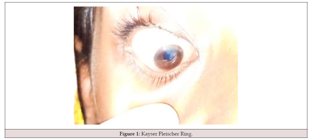
Figure 1:Kayser Fleischer Ring.
Investigation showed normal cell counts in blood picture, Hb 12.3 g/dL; Hct 33.7%; MCV 81.5 fL; MCHC 33.6 g/dL; WBC 8.8 ×10⁹/L; Platelets 295 ×10⁹/L. Normal liver function tests: total bilirubin 0.38 mg/dL; direct bilirubin 0.20 mg/dL; AST 29 U/L; ALT 9 U/L; ALP 260 U/L and renal function tests; urea 8 mg/dL; creatinine 0.9 mg/dL and normal Coagulation: PT 10.7 s (control 14.5 s); PPTT 25.5 s (control 30.0 s); INR 1.02. USG abdomen was normal with no evidence of enlarged liver or spleen. Copper studies showed low serum ceruloplasmin 0.05 g/dL (normal > 0.20 g/ dL); 24-hour urinary copper 271 μg/day (normal < 100 μg/day). The brain imaging showed typical findings of basal ganglia copper deposition suggestive of WD. (shown in figure 2a,b,c) LEIPZIG DIAGNOSTIC score in our patient was 08 (≥4 diagnostic).
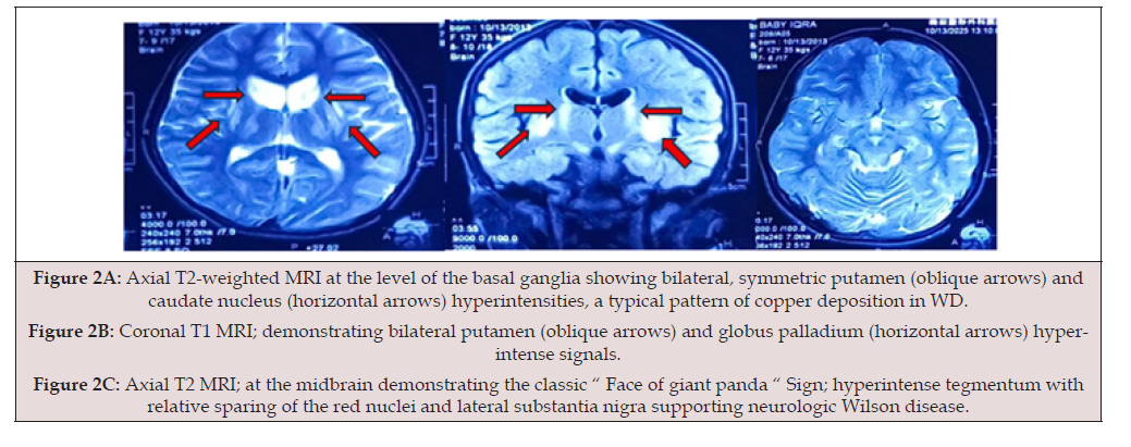
Figure 2A: Axial T2-weighted MRI at the level of the basal ganglia showing bilateral, symmetric putamen (oblique arrows) and caudate nucleus (horizontal arrows) hyperintensities, a typical pattern of copper deposition in WD.
Figure 2B: Coronal T1 MRI; demonstrating bilateral putamen (oblique arrows) and globus palladium (horizontal arrows) hyperintense signals.
Figure 2C: Axial T2 MRI; at the midbrain demonstrating the classic “ Face of giant panda “ Sign; hyperintense tegmentum with relative sparing of the red nuclei and lateral substantia nigra supporting neurologic Wilson disease.
Patient treatment started with restriction of copper containing foods including animal organs (liver, brain, kidneys, heart), Nuts, shellfish, mushrooms and chocolates. Chelation started with D-penicillamine 20mg/kg/day divided x bd with vitamin b6 supplementation. The Zinc acetate started 20 mg x thrice a day prior meal to decrease the gut absorption oof copper and patient kept on follow with monitoring of urine analysis and CBC, to look for d-penicillamine complications. At 3 months of follow up patient had showed significant improvement in gait; he was able to walk with support and choreiform movements resolved. No other medical treatment for dystonia or choreiform movement offered in our patient. The treatment of primary disease reversed the neurologic manifestation in our patient. Our case highlights the very rare < 10% manifestation of WD with only neurologic involvement. Cognition was intact in our patient but exhibited dystonic, choreiform and parkinsonism-like symptoms, which treated with copper chelating agent (Figure 2).
Discussion
Wilson disease is monogenic autosomal recessive disorder. Over 800 mutations are identified to cause the variable disease phenotype. p.H1069Q mutation is most common type (40%) have been shown to have predominantly neurologic presentations. R778L mutation have been shown to have an earlier onset of disease and predominantly hepatic presentation. While p.G943S and p.M769V mutations result in defective copper metabolism but preserved ceruloplasmin levels, which focuses on spectrum of genotype-phenotype relation [4]. Thus, heterogenicity can cause a significant diagnostic challenge, particularly in children Impaired biliary copper excretion and defective incorporation of copper into ceruloplasmin. Progressive accumulation of toxic free copper primarily affects the liver and central nervous system, resulting in a wide range of hepatic, neurological, and psychiatric manifestations [2-4].
Hepatic involvement accounts for approximately 40–60% of initial presentations, particularly in pediatric patients and adolescents. These manifestations range from asymptomatic elevation of transaminases to chronic hepatitis, cirrhosis, and Acute Liver Failure (ALF) [5,6]. Neurological manifestations represent the second most common mode of presentation. Approximately 30–40% of patients present with neurological features, including dystonia, tremor, dysarthria, parkinsonism, ataxia, and gait disturbances. Psychiatric symptoms such as personality changes, depression, and cognitive impairment may accompany or precede overt neurological signs 13 [5-7]. Importantly, a distinct subgroup of patients presents with neurological Wilson’s disease without clinically apparent hepatic involvement. The literature suggests that around 10% of WD patients exhibit isolated neurological manifestations with minimal or no biochemical evidence of liver disease at presentation. However, subclinical hepatic copper accumulation is usually present and may become evident later in the disease course, if not treated with chelation timely [5,7].
Kayser–Fleischer (KF) rings, caused by copper deposition in Descemet’s membrane, are a key diagnostic clue. KF rings are detected in up to 90–98% of patients with neurological WD [8]. Biochemically, low serum ceruloplasmin levels (<20 mg/dL) and elevated 24‑hour urinary copper excretion (>100 μg/24 h) remain central diagnostic hallmarks. Nevertheless, normal ceruloplasmin levels have been reported, particularly in early disease or hepatic‑dominant cases, emphasizing the importance of integrated diagnostic assessment rather than reliance on a single parameter [9,10]. Given the absence of a single definitive diagnostic test, the Leipzig scoring system is widely used to estimate the probability of Wilson’s disease.
Magnetic Resonance Imaging (MRI) plays a crucial role in patients with neurological involvement. Typical MRI findings include symmetrical T2‑weighted hyperintensities in the basal ganglia (particularly the putamen), thalamus, brainstem, and cerebellum. These changes correlate with neurological severity and disease duration and are considered highly supportive of the diagnosis in the appropriate clinical context [5,9,10]. Early diagnosis is critical, as Wilson’s disease is a potentially reversible condition when treated promptly. Chelation therapy with D‑penicillamine or trientine enhances urinary copper excretion, while zinc therapy reduces intestinal copper absorption. Initiation of therapy significantly reverses neurological symptoms, particularly before irreversible tissue damage occurs.
Conclusion
Neurological Wilson disease without overt hepatic involvement requires a high index of suspicion. Early recognition of symptoms and diagnosis is essential for dietary restriction of copper containing foods and early chelation helps to decrease morbidity. Further, family screening and counselling of inheritance pattern is important for next generation.
Author Contribution
Eliya Ali: Manuscript Writing and Literature Review.
Shanti Lal M. Bhojwani: Diagnosis, Conceptualization and
Clinical Management.
Aisha A Haleem: Manuscript Editing and Review.
Kaneez Fatima: Literature Review
Saman: Diagnosis, Manuscript Editing.
Siraj Ahmed: Literature Review
Funding
No funding sources.
Conflict of Interest
None declared.
Ethical Approval
Not required for single case report; patient confidentiality preserved.
Consent
Written informed consent was obtained from the patient/ guardian for publication.
References
- Mak CM, Lam CW (2008) Diagnosis of Wilson's disease: a comprehensive review. Crit Rev Clin Lab Sci 45(3): 263-290
- Porlas RV Jr, de Castillo LLC, Dioquino CPC (2018) Neurologic Wilson disease: case series on a diagnostic and therapeutic emergency. Dialogues Clin Neurosci 20(4): 341-345.
- Lorincz MT (2010) Neurologic Wilson's disease. Ann N Y Acad Sci 1184: 173-187.
- Chang IJ, Hahn SH (2017) The genetics of Wilson disease. Handb Clin Neurol 142: 19-34.
- Dusek P, Litwin T, Członkowska A (2019) Neurologic impairment in Wilson disease. Ann Transl Med 7(Suppl 2): S64.
- Khan Soban, Waqar Zaid, Majid Haris, Farhan Ahmed, Siddique Shajee (2023) A Rare Presentation of Wilson Disease: A Case Report. Pakistan Journal of Neurological Sciences: 18(4).
- Stremmel W, Merle U, Weiskirchen R (2019) Clinical features of Wilson disease. Ann Transl Med 7(Suppl 2): S61.
- Amalnath DS, Subrahmanyam DK (2012) Ocular signs in Wilson disease. Ann Indian Acad Neurol 15(3): 200-201.
- Immergluck J, Grant LM, Anilkumar AC (2026) Wilson Disease. [Updated 2025 Apr 3]. In: StatPearls [Internet], Treasure Island (FL).
- Ortiz JF, Morillo Cox Á, Tambo W, Eskander N, Wirth M, et al. (2020) Neurological Manifestations of Wilson's Disease: Pathophysiology and Localization of Each Component. Cureus 12(11): e11509.




 We use cookies to ensure you get the best experience on our website.