
Review Article 
 Creative Commons, CC-BY
Updates in Etiological Mechanism and Medical Treatment of Hepatic Fibrosis
*Corresponding author:Xukun Deng, School of Pharmaceutical Sciences, South-Central Minzu University, Wuhan, 430074 P. R. China and Xiaoming Wang, State Key Laboratory of Pharmaceutical Biotechnology, Affiliated Drum Tower Hospital, Medical School of Nanjing University, School of Life Sciences, Nanjing University, Nanjing, 210023 P. R. China.
Received:April 06, 2026; Published:April 15, 2026
DOI: 10.34297/AJBSR.2026.30.003978
Abstract
Liver fibrosis is a pathological process triggered by chronic liver injury, characterized by abnormal accumulation of Extracellular Matrix (ECM), which can ultimately lead to cirrhosis and associated complications. During liver injury, Hepatic Stellate Cells (HSCs) become activated and transdifferentiate into myofibroblasts, resulting in an imbalance between ECM deposition and clearance. Without timely intervention, liver fibrosis may progress to cirrhosis, hepatocellular carcinoma, liver failure, or other life-threatening liver diseases. This review summarizes the primary etiological factors contributing to liver fibrosis and discusses the underlying pathogenic mechanisms, with a particular focus on HSC activation, inflammatory responses, and ECM remodelling. Additionally, the review provides an overview of diagnostic approaches for liver fibrosis, including histopathological assessments and non-invasive methods, such as serum biomarkers and advancements in imaging techniques. In terms of treatment and prevention, we highlight recent progress in antifibrotic strategies, including the potential of small molecule drugs and traditional Chinese medicine, and discuss the role of multi-omics research in advancing personalized and precision medicine. The review concludes by addressing key challenges and future directions in liver fibrosis research, particularly regarding the potential applications of personalized and precision therapies, thereby providing critical insights for future research and clinical application.
Keywords:Liver fibrosis, Pathogeny of Hepatic Fibrosis, Pathogenesis of Hepatic Fibrosis, Extracellular Matrix, Antifibrotic Therapy, Omics
Introduction
Hepatic Fibrosis is a pathological component of various chronic liver diseases. It is a repair response to chronic injury and inflammatory wounds, an imbalance between extracellular matrix deposition and clearance, leading to excessive ECM deposition and aggregation [1]. ECM is a non-cellular three-dimensional macromolecular network composed of collagens, elastin, fibronectin, laminins and several other glycoproteins, which regulate diverse cellular functions, such as survival, growth, migration, and differentiation, and are vital for maintaining normal homeostasis [2]. Early Hepatic Fibrosis has a repairing effect on acute liver damage, but late Hepatic Fibrosis can lead to portal hypertension, liver failure, cirrhosis, and even liver cancer [3]. In the pathogenesis of hepatic fibrosis, the activation of HSCs is a significant step in the process of liver fibrosis. When persistent liver damage occurs, HSCs are activated and continue to increase, transforming into myofibroblasts. These cells have been identified as the primary source of collagen production that damages the liver, leading to further aggravation of hepatic fibrosis [4]. Liver fibrosis, characterized by the abnormal accumulation of fibrillar collagen and scar matrix, is a result of dysfunctional wound repair in the presence of chronic liver damage [5]. The global incidence of liver fibrosis is 4.5-9% [6]. Cirrhosis, the end stage of progressive liver fibrosis, is the leading cause of liver-related deaths worldwide. In 2017, deaths due to cirrhosis accounted for 2.4% of total deaths globally and it was the 11th most common cause of death. Although fibrosis is a dynamic disease, it can be reversed if the underlying liver injury is removed or effectively treated [7]. However, advanced cirrhosis is generally irreversible, and there is currently no effective treatment other than hepatic transplantation. Hence, identifying effective and safe treatments for liver fibrosis is of utmost importance. The common causes of hepatic fibrosis are excessive drinking, viral hepatitis, and non-alcoholic fatty liver disease [8], autoimmune and hereditary diseases [9]. Hepatic fibrosis is partly a highly dynamic process [10]. The continuous process of hepatic fibrosis is the late stage of chronic liver disease, which can lead to ascites, jaundice, massive bleeding due to rupture of oesophageal and gastric varices, hepatic encephalopathy, liver cancer, and liver failure [11]. Some studies have shown that the clinical trials of antiviral therapy to block the progress of hepatic fibrosis provide key information for the treatment of hepatic fibrosis, so it is particularly important to actively treat patients early.
Pathogeny of Hepatic Fibrosis
Chronic Viral Hepatitis
Viral hepatitis is an acute inflammation of the liver caused by two sources of infection. It may be a virus named Virus A, which is the cause of infectious or epidemic hepatitis, or Virus B, which is the cause of serum hepatitis or homologous serojaundice. The number of people infected with the hepatitis virus in the world annually is 10 times more than that of AIDS, and the number of deaths due to contracted hepatitis exceeds 1 million every year [12]. Hepatitis A and B viruses can cause acute liver disease, often accompanied by jaundice and systemic symptoms. Hepatitis B and C are at risk of developing cirrhosis and liver cancer. The Hepatitis E virus is easy to cause epidemic diseases in developing countries. Still, it is rare in the United States, and there is no evidence that it can cause acute or chronic liver injury. In HBV and HCV-related liver fibrosis diseases, the general pattern of fibrosis evolution is called post necrosis or bridging fibrosis, which is characterized by the increased deposition of ECM components in the form of portal vein central (vein) fibrosis diaphragm driving or portal vein central bridging necrosis [13]. Primary factors considered in the relationship between HBV or HCV and fibrosis:(1) The injury of liver parenchyma and the death of liver cells should be attributed to the immune response of trying to eliminate HBV and HCV virus. However, the overall response of virus-specific CD4 and CD8 T lymphocytes becomes inefficient over time, and they cannot eliminate HBV or HCV in the liver, leading to chronic infection. (2) The circulation of low-level cell damage and persistent inflammatory reaction, the secondary recruitment of non-antigen specific monocytes (NK cells), which is crucial to the liver parenchyma damage, inflammatory reaction, and fibrosis progress [14]. (3) Direct action by specific HCV proteins such as core and NS3/NS5 proteins. Although HCV does not infect HSCs, these proteins can stimulate activated human HSCs in a ROS and redox-dependent manner, leading to upregulating pro-fibrosis and pro-inflammatory responses [15]. (4) In addition, HCV protein can induce increased ROS production in hepatocytes, which may lead to oxidative stress-mediated hepatocyte injury and activation/ continuation of HSC phenotypic response [16]. Hepatic fibrosis is the initial central stage of viral hepatitis, developing into cirrhosis and liver cancer. Therefore, the focus of diagnosis and treatment of viral hepatitis is to eliminate the virus and prevent it from developing into cirrhosis or liver cancer.
Alcoholic Hepatitis
The normal metabolism of the liver of a person who takes a large amount of alcohol for a long time will be disrupted, which may lead to fatty liver (also known as steatosis), which is a disease that contains large vesicular triglyceride drops in liver cells. Although alcoholic fatty liver can be solved through abstinence, people who continue to drink are prone to liver fibrosis and cirrhosis. Although alcoholic fatty liver can be solved through abstinence, people who continue to drink are prone to liver fibrosis and cirrhosis. Compared with the general population, patients with alcoholic liver disease confirmed by biopsy have a nearly fivefold increased risk of death [17]. In addition, during the 2019 coronavirus disease pandemic, the association between heavy drinking and ALD (autoimmune liver disease) increased, and the consequences of alcohol are expected to continue [18]. It is worth noting that in patients with alcoholic liver disease, macrophages are enriched in the portal vein and can promote ethanol-induced inflammation in vivo, especially in severe alcoholic hepatitis, in which the increase of intestinal permeability and portal vein endotoxin level contributes to the accumulation and activation of macrophages in Ly-6Chi mice and the release of TNF, ROS, etc [19]. In addition, the metabolic pathway of alcohol produces reactive oxygen species, which are effective inducers of lipid peroxidation, leading to necrosis or apoptosis of hepatocytes. Notably, three liver enzymes are involved in the ethanol metabolism to acetaldehyde, which will produce a large amount of NADH during ethanol metabolism. NADH will inhibit the conversion of lactic acid into pyruvate in the process of gluconeogenesis, leading to lactic acid cumulative acidosis and hypoglycemia caused by the inhibition of gluconeogenesis. Acetaldehyde can participate in the modification of many proteins and enzymes in liver tissue. Long time and large amounts of ethanol intake will lead to the accumulation of acetaldehyde in liver tissue, which in the next step will cause functional problems of liver cells and cause damage and death of liver cells. Long-term damage and death of hepatocytes will lead to liver fibrosis and cirrhosis. It can be seen that it is indispensable to control alcohol intake, especially for patients with primary liver diseases such as obesity and viral hepatitis.
Non-alcoholic Fatty Liver Disease
Nonalcoholic Fatty Liver Disease (NAFLD) is liver cell damage, inflammation, and fibrosis caused by liver lipid accumulation, which leads to more severe liver diseases. The global prevalence of NAFLD is 25.24%, with the highest prevalence in the Middle East and South America and the lowest in Africa [20]. Nonalcoholic Fatty Liver Disease (NAFLD) is one of the leading causes of liver cirrhosis in the world [21]. NAFLD is expected to increase exponentially in the next few years, significantly increasing the health system and economic burden [22]. In recent years, there is increasing evidence that NAFLD is a multisystem metabolic disease associated with hyperinsulinemia and genetic susceptibility, with hepatocyte triglyceride accumulation as a major pathologic change. In addition to liver related complications, NAFLD also increases the risk type 2 diabetes mellitus (T2DM), cardiovascular disease and chronic kidney disease. Conversely, obesity, T2DM, lifestyle changes, and drug-resistant genetic alterations also increase NAFLD morbidity and mortality.
Cholestatic Hepatitis
Cholestasis is the obstruction of bile secretion or excretion caused by various reasons, which leads to the inability of bile to flow normally into the duodenum and then back into the blood circulation. Bile transport dysfunction caused by injury of hepatocytes and bile duct cells induces proliferation of bile duct and peritubular myofibroblasts, eventually leading to liver fibrosis. Cholestatic liver disease, such as Primary Biliary Cholangitis (PBC) or Primary Sclerosing Cholangitis (PSC), is a chronic progressive disease, often leading to cirrhosis and subsequent complications. Congenital cholestasis syndrome, whose potential phospholipid invertase (ATP8B1) or bile salt outlet pump (BSEP; ABCB11) dysfunction may also rapidly progress to cirrhosis and often requires liver transplantation [23]. Although the pathogenesis of cholestasis varies, the systemic and hepatic accumulation of hydrophobic bile salts are standard pathogenic features [24]. For example, in PBC, it has been found that late-stage systemic bile salt levels increase by up to 20 times. This widely accepted hypothesis was proposed in the 1970s, and to this day, the accumulation of hydrophobic bile salts is still considered a driving force for fibrosis in cholestatic liver disease [25, 26].
Autoimmune Hepatitis
Autoimmune liver disease includes Autoimmune Hepatitis (AIH), characterized by necrotizing inflammation, primary biliary cirrhosis (PBC), and primary sclerosing cholangitis (PSC), all of which are characterized by progressive cholestasis [27]. Autoimmune hepatitis is an acute or chronic liver parenchymal disease characterized by a loss of tolerance [28] to liver cell-specific autoantigens, which is often caused by environmental and genetic factors [29]. If left untreated, autoimmune hepatitis can usually lead to liver fibrosis, even cirrhosis and liver cancer. Previous studies on patients with autoimmune hepatitis have shown that approximately 30% of patients are diagnosed with cirrhosis. Immunomodulatory therapy can significantly improve the long-term prognosis of patients with autoimmune hepatitis [30]. Monoclonal antibodies targeting key cytokine pathways (monoclonal antibodies targeting CD3[31], CD20[32], and tumour necrosis factor-alpha) can now be used for molecular and cellular interventions. Recombinant molecules, such as cytotoxic T lymphocyte antigen-4 fused with immunoglobulin, may inhibit lymphocyte activation and cellular interventions, such as adoptive transfer of regulatory T cells and sensitization of disease-specific glycolipids to natural killer T cells [33], which may stimulate or inhibit counter-regulatory immune mechanisms. These interventions are now expected to improve treatment outcomes by targeting key areas of immune-mediated pathogenic responses precisely, potentially achieving faster, more complete, and lasting outcomes.
Schistosoma Infected Liver Disease
Liver fibrosis caused by schistosomiasis is essentially a severe parasitic infection caused by the deposition of schistosome eggs in the liver. Due to the host body’s long-term immune response to schistosome egg antigen, hepatic stellate cells are abnormally activated and transformed into MFB, resulting in the deposition of ECM and liver fibrosis after a large number of value-added [34]. Epidemiological studies have shown that schistosomiasis is still the second most common tropical disease. Two hundred forty-nine million people in 73 countries worldwide suffer from different types of schistosomiasis, and nearly 800 million people are threatened by this infection [35]. Although various etiological factors may lead to chronic liver injury leading to liver fibrosis and even cirrhosis, such as drug abuse, alcoholism, and viral infection, schistosomiasis infection is one of the most common causes of liver fibrosis in some countries [36]. Some reports show that effective schistosome treatment could not prevent the development of egg granuloma inflammation and liver fibrosis [37, 38], possibly due to continuous pathological processes such as chronic inflammation. So far, the exact mechanism that mediates the constant activation of inflammation around egg granuloma in the liver during schistosomiasis infection remains unclear.
Pathogenesis of Hepatic Fibrosis
Cells and Hepatic Fibrosis
The liver is composed of parenchymal cells and nonparenchymal cells. Parenchymal cells refer to Hepatocytes (HC), while nonparenchymal cells include Sinusoidal Endothelial Cells (SEC), Kupffer Cells (KC), Hepatic Stellate Cells (HSC), and pit cells. Studies have shown that these cells are involved in developing liver fibrosis in different ways.
Hepatocyte
There are a lot of microvilli on the surface of hepatocytes. During the formation of liver fibrosis, hepatocytes synthesize collagen type Ⅰ, Ⅲ, Ⅳ, Ⅴ, but the amount of collagen synthesis is less than that of HSC. The leading role of HC is to activate HSC. At present, it is believed that the activation mechanism of HC is that the normal hepatocyte membrane has a contact inhibition effect on the proliferation of HSC. In contrast, virus infection, steatosis, or alcohol-induced hepatocyte apoptosis and damage of hepatocyte membrane will lead to the loss of contact inhibition effect on HSC and activate HSC. Apoptotic hepatocytes have been shown to promote the secretion of pro-inflammatory and pro-fibrosis cytokines by macrophages and directly promote HSC activation [39,41]. Recent studies have also demonstrated that inflammatory stressed hepatocyte cytokine (such as IL-33) can promote fibrosis [42].
Hepatic Stellate Cells
HSCs are located in the interspace of the dice around the sinuses and the intercellular fossa of hepatocytes. The cytoplasm is rich in vitamin A-like lipid droplets. HSCs are the central source cells of the Extracellular Matrix (ECM) during liver fibrosis, and their activation is the vital and central event of liver fibrosis [40]. In the fibrotic liver, resting HSC transdifferentiate into proliferative, migratory, and contractile myofibroblasts, which exhibit fibrosis transcription and secretion characteristics (called “cell activation”) and secrete ECM molecules, which accumulate in hepatocytes and form scar tissue. Storage of (retinoid in cytoplasmic droplets is a unique feature of fixed HSC, which is gradually lost during transdifferentiation. However, its causal relationship with HSC activation remains uncertain [39,44]. Endothelin-1 (ET-1) is a potent vasoconstrictor secreted by activated HSC cells that promotes cell proliferation, fibrosis and contraction and may be associated with cirrhotic portal hypertension [43]. Activation includes two main stages: the initiation stage and the sustained stage. The initiation stage refers to the changes in early gene expression and the phenotypic changes in cells produced by cytokines and other stimulating factors. The initial changes in HSC may result from paracrine stimulation from nearby cells, including SEC, HC, KC, etc. During the sustained phase, these stimulating factors maintain the activation phenotype of HSC, resulting in the formation of liver fibrosis. Both autocrine and paracrine pathways regulate the activation of HSC at this stage. As the primary executor of fibrosis, HSC can receive a wide range of signals from injured liver cells and disturbed liver microenvironment. HSC interacts with liver cells, macrophages, lymphocytes, and endothelial cells to promote fibre formation. In addition, activated HSC interacts with natural killer cells, leading to HSC death and termination of fibrosis response. HSC derived from mice or humans exhibit high reactivity to pro-inflammatory cytokines and LPS, activating pro-inflammatory signalling pathways such as nuclear factor-kB and AP-1 and producing chemokines and cytokines (Figure 1). However, compared to the crucial role of macrophages and other white blood cell populations, HSC may have a relatively small contribution to overall liver inflammation. HSC may play a role mainly as receptors for inflammatory signals rather than promoting the overall inflammatory state of the liver [45].
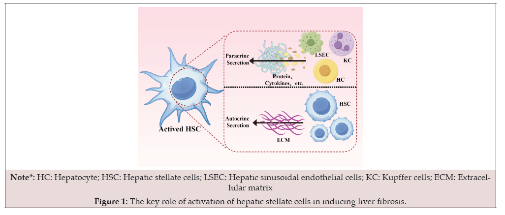
Figure 1:The key role of activation of hepatic stellate cells in inducing liver fibrosis.
Liver Sinusoidal Endothelial Cell
Hepatic Sinusoidal Endothelial Cells (LSEC) are damaged, swollen, and even necrotic, narrowing the hepatic sinuses and leading to the contraction of LSEC and HSC. Expressing adhesion molecules makes inflammatory cells gather in the hepatic sinuses and makes LSEC phenotypic transfer to vascular endothelial cells. Liver fibrosis can be caused by the crosstalk between LSEC and hepatocytes [46]. Hepatocytes and LSEC interact through the VEGFR2 pathway. CD147 is simultaneously expressed in hepatocytes and LSEC. It is a transmembrane glycoprotein associated with liver fibrosis [47] and plays a regulatory role in the information exchange between hepatocytes and LSEC (Figure 2).
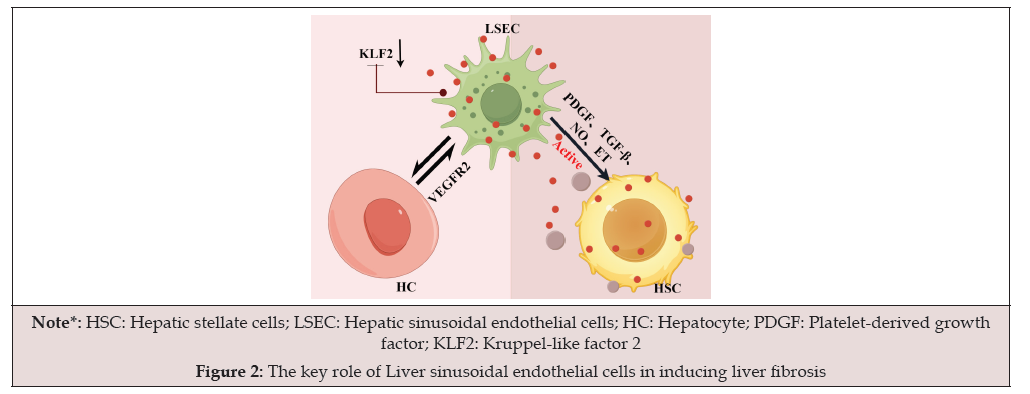
Figure 2:The key role of Liver sinusoidal endothelial cells in inducing liver fibrosis
Interestingly, the anti-CD147 antibody inhibits angiogenesis through the VEGF-a/vegfr2 pathway, thereby improving the progress of liver fibrosis. However, due to the inability to protect adjacent cells, inhibition of hepatocyte apoptosis after injury is not beneficial to preventing liver fibrosis [48]. In addition, the interaction between hepatocytes and LSEC promotes the capillary effect of LSEC, reduces portal vein angiogenesis, and promotes the progress of liver fibrosis. The above effect is achieved by combining leukocyte-derived chemokine 2 (LECT2) expressed by hepatocytes and Tie1 produced by LSEC [49]. Liver fibrosis can also be caused by crosstalk between LSEC and HSC. In continuous liver injury, LSEC is capitalized and has the phenotypes of promoting vasoconstriction, inflammation, angiogenesis, and fibrosis [50]. Capillary LSEC does not promote HSC quiescence but activates HSC by secreting Platelet- Derived Growth Factor (PDGF), TGF-β, and reducing Kruppel-Like Factor 2 (KLF2), a transcription factor that is a protective molecule of liver endothelial vessels. In addition, it has been proved that LSEC-derived fibronectin can also affect the phenotype of HSC and promote its activation [51]. Subsequently, HSC began to increase, contract, and deposit many collagen fibres and extracellular matrix molecules in the liver parenchyma, leading to organ sclerosis and interfering with all cell functions [52].
Interestingly, the anti-CD147 antibody inhibits angiogenesis through the VEGF-a/vegfr2 pathway, thereby improving the progress of liver fibrosis. However, due to the inability to protect adjacent cells, inhibition of hepatocyte apoptosis after injury is not beneficial to preventing liver fibrosis [48]. In addition, the interaction between hepatocytes and LSEC promotes the capillary effect of LSEC, reduces portal vein angiogenesis, and promotes the progress of liver fibrosis. The above effect is achieved by combining leukocyte-derived chemokine 2 (LECT2) expressed by hepatocytes and Tie1 produced by LSEC [49]. Liver fibrosis can also be caused by crosstalk between LSEC and HSC. In continuous liver injury, LSEC is capitalized and has the phenotypes of promoting vasoconstriction, inflammation, angiogenesis, and fibrosis [50]. Capillary LSEC does not promote HSC quiescence but activates HSC by secreting Platelet- Derived Growth Factor (PDGF), TGF-β, and reducing Kruppel-Like Factor 2 (KLF2), a transcription factor that is a protective molecule of liver endothelial vessels. In addition, it has been proved that LSEC-derived fibronectin can also affect the phenotype of HSC and promote its activation [51]. Subsequently, HSC began to increase, contract, and deposit many collagen fibres and extracellular matrix molecules in the liver parenchyma, leading to organ sclerosis and interfering with all cell functions [52]..
Kupffer Cell
Kupffer Cells (KC) are macrophages located in the hepatic sinuses. Their processes can extend into the Disse space under the sinusoidal endothelial cells, sec pores, and direct contact with HC and HSC. With the injury of liver parenchymal cells, the original Kupffer cells in the liver and the monocytes in the blood began to infiltrate and increase into the liver tissue. A KC-conditioned medium can accelerate the activation of HSC in the early stage of culture, increase its proliferation, and promote its secretion of ECM.KC can stimulate the synthesis of extracellular matrix, cell proliferation, and activation of HSC by secreting cytokines, especially TGF-β. TGF-α secreted by KC can induce cell proliferation, while TGF-β secreted by KC stimulates HSC to secrete many ECM. In addition, KC can also affect HSC by secreting matrix metalloproteinase-9 (MMP-9, gelatinase B). MMP-9 can activate TGF-β, which stimulates HSC to secrete collagen. KC is also a significant source of reactive oxygen species intermediates (ROS) in the liver. ROS can promote HSC activation and collagen secretion, whether in or outside the cell. Nitric oxide produced by KC can reduce the proliferation of HSC and inhibit the activation of ROS on HS [54].
Lacunar Cell
Lacunar cells are large granular lymphocytes with natural killer activity in the liver. Their morphological characteristics are cell polarity and aniline blue granules in the cytoplasm. The role of lacuna cells in developing liver fibrosis is unclear. Lacuna cells are significantly increased in the rat model of viral hepatitis. It is speculated that lacuna cells indirectly affect the formation of liver fibrosis by interacting with other non-parenchymal cells (Figure 3).
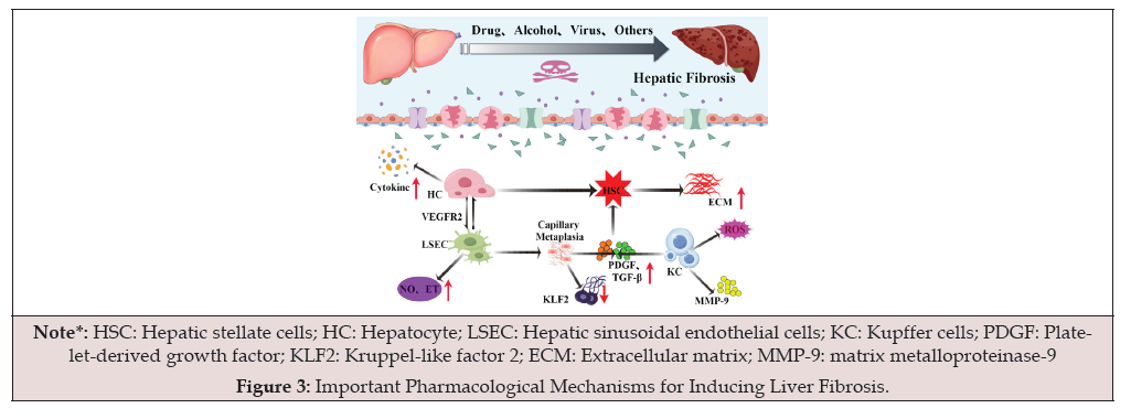
Figure 3:Important Pharmacological Mechanisms for Inducing Liver Fibrosis.
Extracellular Matrix and Hepatic Fibrosis
The Extracellular Matrix (ECM) includes collagen, non-collagen glycoprotein, proteoglycan, and elastin, constituting the matrix of stromal cells and the basement membrane of epithelial cells and blood vessels. During liver fibrosis, the changes in ECM include quantitative changes (excessive collagen formation and deposition in the liver) and qualitative changes (local reconstruction or redistribution of ECM), resulting in the imbalance of ECM synthesis and degradation, excessive accumulation and deposition in the liver, especially the reduction of degradation in the later stage, which is the primary mechanism of liver fibrosis formation. It is believed that the cells that produce matrix in the liver are HC, HSC, SEC, KC, and pit cells, and HSC is the primary source of ECM. In the process of liver injury, HSC is activated and transformed into myofibroblast-like cells, accompanied by increased ECM synthesis and expression of α-SMA. Other ECM components synthesized by HSC include laminin, etc.
Collagen: Collagen protein accounts for 5% -20% of the total protein in the normal liver, but it can increase to about 50% in the case of liver fibrosis. There are several main types of collagen in the case of liver fibrosis, namely, interstitial collagen (type Ⅰ, Ⅲ, Ⅴ collagen), stromal membrane collagen (type Ⅳ collagen), and short chain or microfilament collagen (type Ⅵ collagen), of which type Ⅰ and Ⅲ collagen are the main types. In the normal human liver, type Ⅰ and Ⅲ collagen account for about 80% of the total collagen in the liver. In contrast, in the case of liver fibrosis, type Ⅰ and Ⅲ collagen can account for more than 95% of the total collagen in the liver. Liver fibrosis was mainly type Ⅲ in the early stage and type Ⅰ in the late stage. In the early stage of liver fibrosis, interstitial collagen was deposited in the disease space, which destroyed the functional basement membrane of type Ⅳ collagen. The disease space was capitalized, resulting in blood supply and nutrition supply disorders. In the normal rat liver, the collagen around the hepatic sinuses is composed of newly synthesized type Ⅳ collagen.
In contrast, the portal area mainly comprises mature types Ⅰ and Ⅲ, also essential components of fibrosis. In liver fibrosis, type Ⅰ and Ⅲ collagen is a necessary source of liver fibrous septum. The newly formed type Ⅰ and Ⅲ collagen form fibrous bundles and distribute along these cells in a dendritic manner to create a new fibrous septum. In addition, the collapse of reticular fibrous scaffold caused by cell degeneration and necrosis of mature type Ⅰ and Ⅲ collagen aggravates liver fibrosis. The role of type IV in liver fibrosis is to participate in the capillarization of hepatic sinusoid. Only discontinuous type IV collagen can be seen on the wall of normal hepatic sinusoid. During liver fibrosis, the type IV collagen on the wall of the hepatic sinusoid becomes continuous, and laminin begins to appear, which destroys the functional basement membrane of type IV collagen and forms capillarization. The capillarization of the hepatic sinusoid is one of the foundations for developing liver fibrosis to cirrhosis [55].
Glycoprotein: Non-collagen glycoproteins include Fibronectin (FN), Laminin (LN), and Endokin (EN). FN is a multifunctional macromolecular non-collagenous glycoprotein first found in the body through plasma and cell types. FN can be detected in all the extracellular spaces of the liver. In the early stage of liver fibrosis, plasma FN is suspected to increase, which occurs before the deposition of other extracellular matrix components. Still, this increase is not related to the severity of the disease and other biochemical indicators. It is believed that FN first appears on the fibrous septum and hepatic sinusoid wall in the early stage of liver fibrosis and then is replaced by other interstitial components. The role of FN in liver fibrosis is mainly to play the role of matrix skeleton, conditioning, and cell movement matrix. FN through fibroblasts indicates that adhesion molecules (receptors) will transfer polypeptides into cells, which plays a pacemaker-like role in liver fibrosis. LN is the main glycoprotein in the process of liver fibrosis. Ln mainly exists in the basement membrane and is a unique basement membrane component. LN in the liver comes from HSC, HC, etc. LN and type Ⅳ collagen are distributed in the vascular wall, bile duct wall, and other parts. In the early stage of experimental liver fibrosis, the rough endoplasmic reticulum of HSC was dilated, and there was apparent ln staining, suggesting that the cells synthesized a large amount of LN. In the late stage of liver cirrhosis, the intense positive staining on the sinusoidal wall was distributed with type Ⅳ collagen, indicating that both were involved in liver sinusoidal capillarization. Electron microscopy also confirmed a specific binding point between LN and type Ⅳ collagen. Thus, LN is involved in the formation of liver fibrosis. The role of LN in liver fibrosis is mainly to connect the macromolecular components of the matrix and jointly participate in the formation of the basement membrane and the capillarization of the hepatic sinusoid. Ln can crosslink with type Ⅳ collagen, non-collagen glycoprotein, heparin sulfate, and other matrix components and can crosslink itself, which plays a vital role in forming basement membrane.
Matrix Metalloproteinases and Their Inhibitors: ECM degradation is mainly mediated by Matrix Metalloproteinases (MMPs). Studies have shown that in the early stage of liver fibrosis, proenzyme activity changes little, and the rapid degradation system mainly degrades the increased collagen synthesis at this stage in cells. In the process of fibrosis development, the activity of MMP gradually increased, and in the late stage of liver fibrosis, the activity gradually decreased. Further studies showed that the activity of MMPs could be inhibited by Tissue Inhibitors of Metalloproteinases (TIMPs), which were specific inhibitors of MMPs. TIMPs were the corresponding side regulators of MMPs in the metabolic regulation of ECM and were synthesized and secreted in the same cells as MMPs. TIMPs combine with active MMPs to form a 1:1 chemical complex, which specifically inhibits the activity of MMPs and promotes cell proliferation. The main effect is to inhibit the activation of MMP zymogen and its degradation of EC [56].
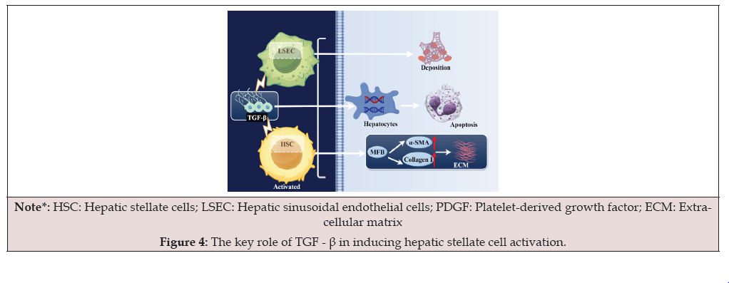
Figure 4:The key role of TGF - β in inducing hepatic stellate cell activation.
Insulin-like Growth Factor
Insulin-like Growth Factor (IGF) is a mitogen-like peptide in cells. There are three peptide hormones in the IGFs family, Namely Insulin (INS), IGF-Ⅰ, and IGF-Ⅱ, which bind to IGFs binding proteins and regulate the role of IGFs through IGFBP.IGFs can promote cell proliferation, differentiation, and maturation, inhibit cell apoptosis, mediate most of the effects of growth hormone, promote growth and anabolism, reduce blood glucose, and regulate immunity. During the recovery period of liver fibrosis, activated HSCs are reduced mainly through apoptosis rather than phenotypic transformation. Cell growth depends on various growth factors. IGF and its receptors are the central regulators of inhibiting cell apoptosis. Experiments have corroborated that IGF-I can significantly inhibit the apoptosis of cycloheximide-induced HSC. In vitro experiments showed that PDGF could substantially enhance the secretion and release of IGF-1 and IGF-1 binding protein in rat hepatic stellate cells and strengthen the activity of IGF-1 by increasing the binding with IGF-1. IGF-1 and PDGF jointly stimulated the DNA synthesis in rat hepatic stellate cells and promoted their division and proliferation, suggesting that the interaction between IGF-1 and other cytokines, such as PDGF, contributes to the formation of liver fibrosis.
Tumor Necrosis Factor Alpha
Tumour necrosis factor-α (TNF-α) is mainly produced by monocyte macrophages, HSC, Kupffer cells, and so on, which have pro-inflammatory activities and cytotoxic effects. In the process of liver fibrosis, TNF-α plays an essential role in the proliferation and activation of HSC, the synthesis of ECM, and the release of matrix metalloproteinases and tissue inhibitors. Studies have found that TNF-α can enhance the proliferation of HSC, promote the transformation of HSC to MFB, and increase the effect of TGF-β on the synthesis of ECM. However, some scholars directly intervened in primary cultured HSCs with TNF-α and found that TNF-α inhibited the proliferation and apoptosis of HSC and induced the “activation” of HSC. The effect of TNF-α on the synthesis of the HSC matrix occurs at the gene transcription stage. The results showed that TNF-α could induce apoptosis of HSC, which was related to its activation state. Its role in promoting HSC apoptosis may be linked to inhibiting the expression of Bcl-2, promoting the expression of Bax, and down-regulating the value of Bcl-2/Bax. In a word, the effect of TNF-α on HSC is multifaceted, and the mechanism of action is complex, which not only promotes liver fibrosis but also inhibits liver fibrosis. TNF-α and other cytokines such as TGF-β, PDGF, and IL-1 form a regulatory network vital in initiating and regulating liver fibrosis.
Angiotensin Ⅱ
Angiotensin Ⅱ (Ang Ⅱ) is the primary bioactive substance secreted by the Renin-Angiotensin System (RAS). Ang Ⅱ can exert its biological effects only by binding to specific receptors on tissue cell membranes, mainly angiotensin Ⅱ type 1 receptor (AT1R). Active hepatic stellate cells have local RAS. The effects on activated hepatic stellate cells include: (1) Promote the activation and proliferation of hepatic stellate cells. Studies have reported that exogenous and Ⅱ can induce a dose-dependent increase of intracellular Ca2+concentration in cultured human hepatic stellate cells mainly through L-type calcium channels, and the increase of intracellular Ca2+concentration is an essential condition for the activation and proliferation of hepatic stellate cells. (2) Promote the contraction of hepatic stellate cells. Studies have shown that Ang Ⅱ can induce the contraction of hepatic stellate cells in a dose-dependent manner, which is closely related to the change of intracellular Ca2+ concentration. The contraction intensity was approximately equal to that of ET-1 and could be inhibited by losartan, suggesting that AT1R mediated this effect. (3) Promote the synthesis of ECM by hepatic stellate cells. Studies have found that Ang Ⅱ can promote the synthesis of collagen and the secretion of hyaluronic acid and laminin in hepatic stellate cells in a dosedependent manner.
It is speculated that Ang Ⅱ may activate a series of signal transduction pathways by binding to AT1R on the membrane of hepatic stellate cells and increasing the expression of TGF-β 1 and PDGF, thereby stimulating the synthesis of collagen in hepatic stellate cells. (4) Inhibit the degradation of ECM by hepatic stellate cells. It was found that Ang Ⅱ could promote the production of TIMP-1 in activated rat hepatic stellate cells in a dose and timedependent manner. Ang Ⅱ binds to AT1R on the membrane of hepatic stellate cells and promotes TIMP-1 production by activating the protein kinase C (PKC) signalling pathway, which AT1R antagonists and PKC inhibitors can inhibit. (5) It is involved in the network regulation of cytokines by hepatic stellate cells. In cultured activated hepatic stellate cells, Ⅱ can induce the expression of TGF-β 1mrna through AT1R, which can be completely inhibited by candesartan, an AT1R antagonist. (6) Inhibition of hepatic stellate cells. Studies showed that Ang-II upregulated Bcl-2 expression but not Bax expression, increased Bcl-2/Bax ratio and inhibited apoptosis of hepatic stellate cells.
Interleukin
Interleukin-1 (IL-1) comes from KC and sec. Its particular biological activity is to promote the proliferation of fibroblasts and HSCs, increase collagen synthesis, increase ECM production, and inhibit MMP synthesis. It is involved in the process of liver fibrosis in many links. Interleukin-6 (IL-6) is also mainly secreted by KC. Activated HSC cells and myofibroblasts are also the primary sources of IL-6, which can stimulate the proliferation of HSC, induce the production of a variety of acute phase proteins, and promote the deposition of ECM by promoting matrix degeneration or interacting with its adhesion receptors. Recent studies have shown that interleukin-10 (IL-10) may be an anti-hepatic fibrosis factor. Active HSC can negatively regulate liver fibrosis by autocrine IL-10, inhibiting type I collagen transcription and stimulating collagenase production.
Platelet Activating Factor
Platelet-Activating Factor (PAF) belongs to phospholipids, which can regulate inflammation, vasoconstriction, and other physiological functions. PAF mediates the interaction between cells and causes the change in cell phenotype. It is speculated that PAF may also be involved in the activation of HSC. Studies have found that KC and HSC can secrete PAF in the early stage of acute liver injury, and Ca2+ carriers, thrombin, and LPS can stimulate the secretion of PAF. Studies have shown that PAF is the primary inflammatory mediator secreted by HSC after liver injury by carbon tetrachloride or free radicals.
Leptin
Studies have found that HSC isolated in vitro and in vivo can produce leptin during activation. In the animal model of chronic liver injury induced by fatty liver and carbon tetrachloride, it was found that leptin-deficient rats had no liver fibrosis and typical liver structure. Liver endothelial cells and Kupffer cells are the primary target cells of leptin-induced liver fibrosis. Leptin can increase the expression of type I procollagen mRNA and TGF-β type II receptor mRNA and protein, enhance the effect of TGF-β 1 on collagen synthesis, and enhance the proliferation of PDGF-dependent HSCs through the PI3K/Akt pathway.
Activin
Activin (ACT) is a member of the large family of TGF β peptide factors, but it cannot bind to the receptor of TGF-β. HSC in resting state does not express ACT, but activated HSC does. In liver fibrosis, HSC expresses a large amount of ACT. There are act-a receptors on the surface of HSC. Smad Protein is vital in act-a signal transduction from cell surface silk and threonine kinase receptors to nuclear target genes. ACT expression levels of HSC at different locations in the hepatic lobule. The closer the hepatic lobule is to the fibrous connective tissue, the higher the gap expression level, showing a gradient distribution. ACT can promote HSC to secrete type I collagen, fibronectin, ECM, and so on in a dose-dependent manner within a specific concentration range, and act-a can play this biological effect in coordination with TGF-β. HSC secreting a large amount of act can also cause apoptosis of surrounding hepatocytes.
Endothelin
Endothelin-1 (ET-1) has a contractile solid effect on HSC. Activated HSC expresses the ET-1 receptor, and the binding of ET-1 with the receptor can increase the content of intracellular Ca2+, causing the contraction of myosin and sarcoplasmic protein. It also increases the expression of α-SMA and regulates the proliferation of HSC. An increase in intracellular Ca2+ concentration and cell contraction induced by ET-1 was observed at any stage of HSC activation. PDGF, TGF-β antagonists, and oxygen stress can increase the release of ET-1 from HSCs. ET-1 acts directly on the hepatic sinusoids, resulting in a decrease in portal vein blood flow in the body.
Epidermal Growth Factor
HSCs proliferate, and epidermal growth factor-r (EGF-R) expression increases in hepatic fibrosis. These findings suggest that EGF has a positive regulatory effect on the gene expression of MMP- 3 and TIMP-1 in HSC and may be a synergistic regulation. The role of EGF in the occurrence and development of liver fibrosis depends on the effect of EGF on MMPs and TIMPs, the interaction between EGF and other cytokines, and the role of EGF in the balance of cytokines.
Diagnosis of Hepatic Fibrosis
Diagnosis of Hepatic Fibrosis
Basic requirements for histopathological examination of liver biopsy: case histological examination is an essential basis for precise diagnosis, measurement of inflammatory activity, degree of fibrosis, and determination of drug efficacy. The liver biopsy specimens should be made into serial sections and routinely stained with hematoxylin-eosin, reticular fibres, and Masson trichrome to accurately judge the degree of inflammation, structural changes, and fibrosis in the liver. Immunohistochemical staining or in situ, viral antigen, or nucleic acid examination were added.
Noninvasive Diagnosis
Serum Markers: They should be relevant parameters that help to predict or detect inflammation and fibrosis in the liver, liver fibrogenesis, or degradation. At present, it is considered that hyaluronic acid (HA), type Ⅲ procollagen peptide or its metabolic fragments (including P Ⅲ P, P Ⅲ NP, P Ⅲ CP), type Ⅳ collagen or its metabolic fragments (including P Ⅳ-NP, P Ⅳ-NC1, P Ⅳ) and laminin (LN) which react to Extracellular Matrix (ECM) components; The combined detection of matrix protease inhibitor-1 (TIMP-1), which reflects the changes of ECM related enzymes, and transforming growth factor β 1 (TGF β1), which demonstrates the formation of fibrosis, is more significant. Two or more of the above six indicators are abnormal, which has the diagnostic significance of liver fibrosis. Related Liver Function and Immune Function: In addition to child Pugh classification, albumin, prothrombin time, aspartate aminotransferase, chlorate aminotransferase, γ-glutamyltranspeptidase, apolipoprotein A1, α2-macroglobulin, γ-globulin, 1GG, and alpha-fetoprotein can be used for auxiliary observation.
Imaging Evaluation: The reasonable selection of ultrasound, CT, and MRI and their mutual verification are helpful for dynamic observation. Quantitative or semi-quantitative criteria were used to observe the changes in liver elasticity, liver volume, liver surface edge, liver abdominal thickness, liver parenchyma, intrahepatic blood vessels, bile ducts, spleen, and splenic vein, gallbladder, and other indicators, which can provide valuable reference data for the diagnosis of liver fibrosis and the evaluation of the activity of the lesions. Transient elastography is also a standard imaging evaluation method. Transient Elastography (TE) is a non-invasive method specially used to measure liver fibrosis. The device has a probe that can emit ultrasound and an elastic wave that passes through the liver parenchyma, which measures Liver Stiffness (LS). Ultrasound (US) is usually the first-line radiological examination for patients with suspected liver cirrhosis because it is easy to obtain, noninvasive, well tolerated, cheaper than CT or MRI peers, provides real-time image acquisition and display, and will not expose patients to adverse reactions of intravenous contrast agent or radiation. The software on the machine processes the crossvelocity data of elastic waves in the liver and provides the value in kilopascals for each measurement. The median of 10 measurements is the examination result. Since 2005, TE has entered the clinical practice of Hepatology, especially in evaluating patients with chronic viral hepatitis [59]. In 2015, the operation was also used to select hepatitis C patients with severe fibrosis and cirrhosis for new antiviral treatment [60].
Treatment of Liver Fibrosis
Liver fibrosis is potentially reversible. Patients receiving treatment for HCV infection experience significant reversal of fibrosis after achieving complete HCV negativity, but this improvement is mainly seen in the early stages (fibrosis grades 1 and 2). However, once fibrosis progresses to more advanced stages (grades 3 and 4) with the formation of cross-linked type I collagen, often accompanied by cell damage and inflammation, this reversibility becomes limite [13]. Therefore, addressing liver fibrosis is both a necessary and urgent task.
Research on the treatment of liver fibrosis has been steadily progressing. Current therapeutic approaches are primarily categorized into Western medicine and traditional Chinese medicine. Western medicine predominantly involves smallmolecule chemical drugs, focusing on etiological treatment and anti-fibrotic therapy. Conventional Chinese medicine mainly utilizes herbal medicines to inhibit liver fibrosis.
Small Molecule Chemical Therapy
Etiological Treatment
The etiological treatment effectively prevents liver damage and is a viable method for treating liver fibrosis. In patients with chronic hepatitis C, significant fibrosis regression is observed following treatment with pegylated interferon α-2a or α-2b combined with ribavirin, particularly in mild to moderate cases, with women showing a higher degree of fibrosis reduction. For alcoholic hepatitis, complete abstinence from alcohol is essential. In many mild cases, avoiding alcohol can resolve clinical symptoms. For non-alcoholic hepatitis, lifestyle improvements through diet and exercise, as well as weight loss surgery, are the most effective methods [61]. For cholestatic hepatitis, Ursodeoxycholic Acid (UDCA) is widely used as a first-line treatment, and bile acid inhibitors, anti-fibrotic drugs, and anti-inflammatory drugs can also be utilized [62]. Continuous corticosteroid therapy can improve autoimmune hepatitis [63]. For patients with advanced liver disease, the limited availability of approved drugs necessitates liver transplantation as the final option for advanced fibrosis [64]. Liver fibrosis caused by schistosomiasis (Schistosoma mansoni and Schistosoma japonicum) is currently treated with praziquantel, while several urgently needed vaccines are in various stages of clinical development and are not yet approved for public use [65]. Etiological treatment is crucial for different causes of liver fibrosis to achieve optimal therapeutic outcomes.
Anti-fibrotic Treatment
For patients with advanced cirrhosis, etiological treatment is relatively slow in effectiveness, necessitating the development of anti-fibrotic treatments with fewer side effects. To date, many antifibrotic therapies have shown success in trials and experiments. These therapies include growth factors, cytokines, miRNA, monoclonal antibodies, stem cell-based methods, and other ECM-targeting approaches. The results of preclinical and clinical studies have laid the foundation for future alternatives to liver transplantation [66].
Activated HSCs, portal vein myofibroblasts, and the ECM they produce play pivotal roles in fibrosis and are thus the primary focus of anti-fibrotic therapies. These cells not only drive fibrotic processes but also interact with other cell types to influence fibrosis dynamics. For instance, they can promote fibrotic activation, induce quiescence or apoptosis, and contribute to ECM remodeling through the release of fibrinolytic enzymes and phagocytosis. This complexity highlights the potential for diverse therapeutic strategies. Therapies directly targeting HSCs, myofibroblasts, and ECM are categorized as “direct anti-fibrotic drugs,” while those modulating other cellular interactions or pathways, such as immune cells or inflammatory mediators, are classified as “indirect anti-fibrotic drugs [67].”
Inhibition of the TGF-β1/Smad Signaling Pathway
Extensive research has shown that dysregulation of the TGF-β1/ Smad pathway plays a crucial role in the development of tissue fibrosis. Smad2 and Smad3 serve as major downstream mediators that facilitate TGF-β1-driven fibrosis, while Smad7 functions as a negative feedback regulator, providing protection against fibrosis induced by TGF-β1.[68]. Therefore, inhibiting the TGF-β1/Smad signalling pathway prevents HSC activation and proliferation, thereby improving liver fibrosis [69]. Experimented by Leonel et al., It is revealed that pirfenidone treatment in animal livers significantly downregulated TGF-β1. It was concluded that pirfenidone might inhibit TGF-β1expression, leading to upregulation of the Smad- 7 gene, which could benefit the treatment of human liver fibrosis [70]. Drugs such as praziquantel and fluorofenidone (Figure 5) also inhibit hepatic stellate cell activation by targeting the TGF-β1/ Smad pathway, serving as useful anti-fibrotic agents [71, 72].
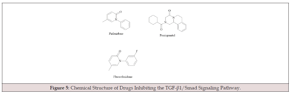
Figure 5:Chemical Structure of Drugs Inhibiting the TGF-β1/Smad Signaling Pathway.
Inhibition of Fibroblast Growth Factors
There are 22 known members of the human Fibroblast Growth Factor (FGF) family. With several physiological roles, FGF21 is an endocrine hormone that is expressed in the liver, pancreas, white adipose tissue (WAT), and brown adipose tissue (BAT) [73]. FGF21 is a crucial metabolic regulator predominantly expressed in the liver. Cong evaluated the effects of the FGF21 analogue LY2405319 in a mouse model of liver fibrosis and concluded that FGF21, as an inhibitor of the succinate-GPR91 pathway, effectively controls liver fibrosis [74]. Thus, FGF21 presents a novel approach for treating fibrosis. Pegbelfermin (BMS-986036), a PEGylated FGF21 analogue, demonstrated in a multicenter, randomized, placebocontrolled phase 2a study that daily doses of 10 mg or weekly doses of 20 mg for 16 weeks were generally well tolerated and considerly reduced liver fat in patients with non-alcoholic steatohepatitis (fibrosis stages 1-3). It also positively affected several parameters related to non-alcoholic steatohepatitis. Furthermore, Harrison et al. found that NGM282, an FGF19 analogue, inhibits classical bile acid synthesis and suppresses fatty acid synthesis and lipogenesis, effectively addressing non-alcoholic steatohepatitis and inhibiting liver fibrosis [75].
Inhibition of ECM
ECM buildup is a hallmark of liver fibrosis, a wound-healing reaction to persistent liver injury. Urokinase Plasminogen Activator (UPA) promotes ECM degradation by activating Matrix Metalloproteinases (MMPs). Increased MMP-9 activity contributes to liver fibrosis, while FRETOH downregulates UPA, MMP-2, and MMP-9 activities in hepatocytes and reduces MMP-9 activity [76]. Thus, FRETOH demonstrates a therapeutic effect on liver fibrosis.
Inhibition of LOX
A humanized IgG4 monoclonal antibody named simtuzumab specifically targets LOXL2, blocking its function in vitro and in mouse studies. In mouse models, simtuzumab reduces fibroblast activation and the production of cytokines and growth factors, such as transforming growth factor-β (TGF-β) [77]. However, further research is required to investigate Simtuzumab’s effect on transaminases, as current experiments do not provide sufficient evidence of its efficacy.
MiRNA and SiRNA
Hepatic stellate cells (HSCs) and fibroblasts, the main source of fibrotic cells in liver tissue, have significant levels of MiR-122 expression. MiR-122 suppresses the expression of serum response factor (SRF), which in turn lowers the transcription of α-SMA and COL1A1, and directly inhibits FN1 expression by binding to its 3’-untranslated region. SRF is an essential transcription factor that mediates fibrotic cell activation [78]. Li demonstrated through lossof- function analysis that the deletion of HDAC2 in TGF-β1 activated HSC-T6 cells promotes cell cycle arrest and blocks the expression of Col1α1 and α-SMA proteins [79]. Therefore, HDAC2 plays a crucial role in HSC activation and liver fibrosis and could be a target for liver fibrosis therapy, although its regulatory role requires further investigation.
Renin-Angiotensin Inhibitors
The renin-angiotensin system (RAS) is an essential regulator of liver cirrhosis and portal hypertension. As liver fibrosis progresses, the levels of RAS components, including angiotensin (Ang) II, Ang- (1-7), Angiotensin-Converting Enzyme (ACE), and Ang II Type 1 Receptor (AT1R), increase [80]. The fibrotic and inflammatory effects of Angiotensin II in the liver are primarily mediated by the Angiotensin Type 1 (AT1) receptor. AT1 receptor blockers have been proposed for treating liver fibrosis in patients with chronic liver diseases and have shown good tolerance. Losartan can reduce serum transaminase levels without adverse effects as a representative drug. Valsartan and Irbesartan (Figure 6) are also commonly used AT1 receptor blockers, showing potential for treating liver fibrosis.
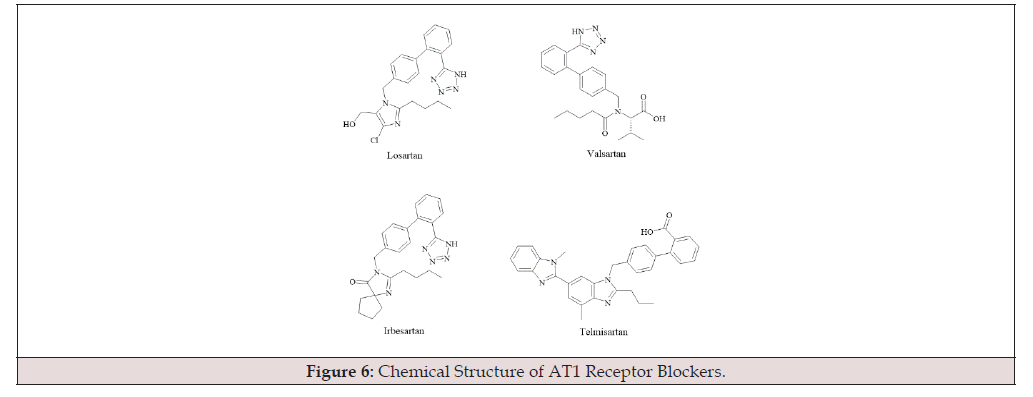
Figure 6:Chemical Structure of AT1 Receptor Blockers.
FXR Receptor Inhibitors
Obeticholic Acid (OCA) is an FXR receptor agonist with an efficacy 100 times greater than Chenodeoxycholic Acid (CDCA). FXR signalling protects hepatocytes from bile acid toxicity by inhibiting bile acid synthesis and stimulating bile secretion through the upregulation of bile acid transporters. Additionally, it regulates other pathways with direct anti-inflammatory and antifibrotic effects. In a clinical trial, OCA improved liver biochemistry [81]. Glycochenodeoxycholic Acid (GUDCA) and Tauroursodeoxycholic acid (TUDCA) (Figure 7) are FXR antagonists that significantly inhibit FXR transcriptional activity.
C-C Chemokine Antagonists
Cenicriviroc (Figure 8), a C-C Chemokine Receptor Type 2 and 5 (CCR2/CCR5) antagonist, has been demonstrated to have antiinflammatory and antifibrotic effects in animal models, which is primarily due to cenicriviroc’s ability to block the inflammatory cascade of fibrosis. Compared to placebo, cenicriviroc significantly improved liver fibrosis by at least one stage after 48 weeks [82].
GR-MD-02
GR-MD-02 (Galactose arabinose-rhamnose cytathiate) has been shown to reduce liver fibrosis and decrease serum biomarkers of NASH fibrosis in animal studies. However, it was found that the drug only had a positive therapeutic effect on a small portion of liver disease patients due to insufficient dosage or lack of sensitivity to high fibrosis levels in experiments [83]. Further clinical trials are needed to determine its efficacy.
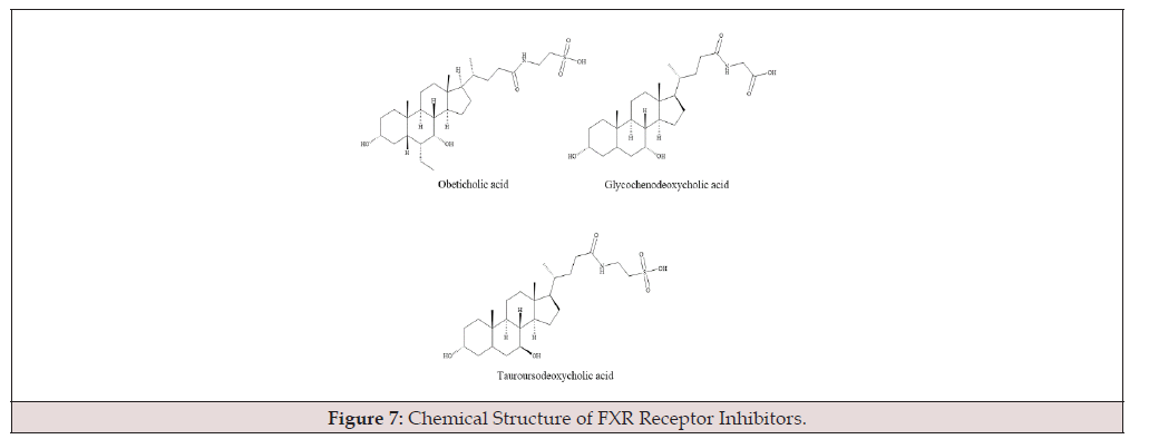
Figure 7:Chemical Structure of FXR Receptor Inhibitors.

Figure 8:Chemical structure of cenicriviroc.
T2DM
In the general population, Type 2 Diabetes Mellitus (T2DM) has been found to be a risk factor for liver fibrosis, particularly in individuals with fatty liver disease. It has also been discovered that metformin, a first-line hypoglycemic medication, helps with liver fibrosis and steatosis [84]. According to liver biopsy results, 32% of patients exhibited advanced fibrosis, and the risk of advanced fibrosis significantly decreased after metformin treatment. Studies have shown that metformin (Figure 9) inhibits liver fibrosis, reduces fibrosis severity, and curtails the progression of non-alcoholic fatty liver disease. Additionally, metformin intervention can inhibit the activation of HSCs, deplete lipid accumulation in hepatocytes, and prevent decompensated cirrhosis and liver fibrosis [85].

Figure 9:Chemical structure of metformin.
Targeting Reactive Oxygen Species (ROS) in Therapeutic Interventions
Research has identified ROS-generating enzymes in many molecules and subcellular organelles, which produce ROS during physiological processes. These ROS-producing enzymes are promising targets for treating oxidative stress-related diseases, including liver fibrosis. For example, NOX and TLR in the liver can generate ROS through crosstalk, potentially adversely affecting liver cell function and leading to fibrosis. Drugs targeting ROS production have received significant attention in treating oxidative stress-related diseases, especially liver diseases, providing adequate means for current clinical therapies [86].
NOX Inhibitors
Reactive Oxygen Species (ROS) generated by NADPH Oxidase (NOX) play a critical role in liver injury and fibrosis. Therefore, inhibiting NOX1 and NOX4 is crucial for treating liver fibrosis. The dual NOX1/4 inhibitor GKT137831 has been shown to inhibit ROS production and inflammation and proliferation genes in primary mouse HSCs. Clinical trials have found that it effectively reduces the degree of liver fibrosis and exhibits good pharmacological properties and safety [87], making it a novel therapeutic approach. Losartan, another drug affecting NOX, is widely used in clinical practice; however, its therapeutic effects on liver-related diseases are not yet significant [86]. NOX1 and NOX4 provide new directions for future research in liver fibrosis treatment.
Antioxidants
Antioxidants can reduce ROS production, thereby improving liver fibrosis. Antioxidants such as S-Adenosylmethionine (SAMe), Silymarin, Lecithin, N-Acetylcysteine (NAC), and Vitamin E (Figure 10) have been proven to have beneficial effects in clinical trials [88].
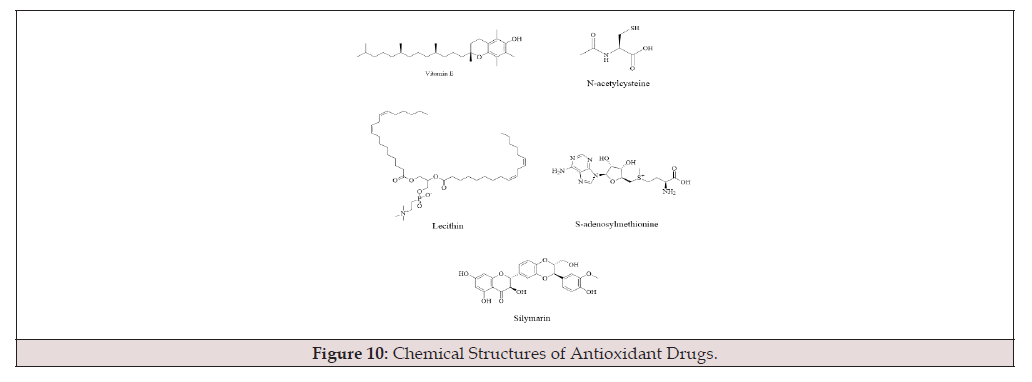
Figure 10:Chemical Structures of Antioxidant Drugs.
Traditional Chinese Medicine (TCM) in Treatment
In recent years, TCM has continuously developed, and its efficacy and safety in the clinical treatment of liver fibrosis have been proven to surpass those of Western medicine, demonstrating a promising application prospect. Unlike Western medicine, TCM emphasizes unity, considering the patient and environmental factors together and treating the patient as an organic whole. TCM exhibits significant biological advantages [89]. The metabolism of intestinal microorganisms affects the therapeutic effect of TCM and is an essential factor to consider.
The liver and intestines are inseparable in the human body. Endotoxemia in liver cirrhosis is a phenomenon of bacterial translocation caused by bacterial overgrowth. Due to the interaction between the intestines and the liver, liver fibrosis affects the intestinal flora [90]. Similarly, the disruption of the gut microbiota can lead to the development of liver fibrosis. It has been found that TCM can effectively regulate the gut microbiota, playing a therapeutic role [91].
Scutellaria Baicalensis
Probiotics can improve the intestinal barrier and protect it from damage. Studies have found that probiotics such as Lactobacillus casei GKC1 (GKC1), Bifidobacterium lactis GKK2 (GKK2), Lactobacillus rhamnosus GKLC1 (GKLC1), and Lactobacillus paracasei GKS6 (GKS6) have potential benefits in alleviating liver fibrosis [92]. Scutellaria baicalensis, a traditional Chinese herb widely used in China for its anti-inflammatory, antibacterial, and antioxidant properties, can effectively promote the healthy growth of intestinal bacteria, making it helpful in treating liver fibrosis [91]. Oroxylin A in Scutellaria baicalensis can induce autophagy of ferritin, thereby inducing HSC senescence and significantly inhibiting the expression of senescence marker SA-β-gal in HSCs, reducing liver fibrosis [93].
Astragalus
Astragalus polysaccharide, an important active component extracted from Astragalus, has antioxidant, antitumor, antiviral, and cardiovascular protective effects and is widely used in the clinical treatment of colitis. IL-17 has strong pro-inflammatory effects, leading to inflammation, while Astragalus polysaccharide can inhibit IL-17 and TNF-α, acting as an anti-inflammatory agent [92]. Astragalus polysaccharide also increases the number of Lactobacillus and Bifidobacterium, further inhibiting inflammation. Serum ALT and AST levels are essential indicators of liver function; Astragalus polysaccharide can significantly reduce serum ALT and AST levels, further indicating its potential to reduce liver disease and protect the liver [94].
Atractylodes
Atractylodes is a traditional Chinese medicine with spleenstrengthening and antidiarrheal effects. Treatment with Atractylodes significantly reduces the number of pathogenic bacteria, such as Helicobacter pylori and Clostridium, while increasing beneficial bacteria, such as Lactobacillus. This indicates that Atractylodes can promote the growth of beneficial bacteria, restore intestinal microecology, and improve intestinal structure, having a therapeutic effect on liver fibrosis [95].
Pueraria
Pueraria, the dried root of the leguminous plant Pueraria, contains many bioactive components with antioxidant, antiinflammatory, and immunomodulatory activities [96]. Research has found that Pueraria extract (RPE) can reduce pathological changes and lipid deposition in rat liver tissue and decrease serum ALT, AST, and liver GGT activity, effectively mitigating liver damage caused by chronic alcohol intake [97]. Animal experiments have shown that puerarin, an active ingredient in Pueraria, activates endogenous PPAR-γ expression in rats, inhibiting the proliferation and activation of ECM-driven liver cells and strongly inhibiting fibrosis [98]. Pueraria and silymarin have therapeutic effects on alcoholic fatty liver and liver inflammation, showing potential as effective drugs for treating liver fibrosis [99].
Schisandra
Schisandra regulates the gut microbiota, reversing the intestinal bile acid spectrum and microbiome to protect the liver and block fibrosis [100]. Schisandra acidic polysaccharides, one of the main components of Schisandra, improve damaged liver tissues pathologically and have an excellent protective effect on acute ethanol-induced liver injury. The mechanism may be related to reducing oxidative stress and regulating the expression of CYP2E1[101]. Schisandra is a natural product with promising future research potential for developing liver-protective drugs.
Sophora Flavescens and Ligustrum Lucidum
Sophora root is an essential herb in treating hepatitis B in China, with its alkaloids showing significant liver-protective effects and polysaccharides demonstrating good immunomodulatory activity [102]. Ligustrum lucidum, the mature fruit of the Oleaceae family, has multiple pharmacological effects, including anticancer, liver protection, anti-inflammatory, anti-osteoporosis, and antioxidant properties [103]. The combination of Sophora and Ligustrum has been documented in ancient medical texts. Studies have found that when the ratio of these two drugs is less than 1:1, the synergistic therapeutic effect on liver fibrosis is significantly enhanced [104]. In the treatment of liver fibrosis, traditional therapies and herbal medicine demonstrate distinct advantages and characteristics. Traditional therapies typically target HSC activation, regulate inflammatory responses, and degrade ECM through mechanisms such as those employed by drugs like lovastatin, which exhibit significant efficacy in inhibiting fibrotic signaling pathways. However, traditional methods also have limitations, including drug resistance, immunosuppression, and potential side effects. In contrast, herbal medicine stands out with its multi-targeted mechanisms, not only modulating HSC activation but also enhancing liver repair through anti-inflammatory, antioxidant, and gut microbiota-regulating pathways. For instance, Scutellaria baicalensis contains flavonoid compounds that exert anti-inflammatory and anti-fibrotic effects by inhibiting the TGF-β signaling pathway; Atractylodes macrocephaly has been shown to improve liver function, modulate the immune system, and reduce ECM deposition; while Pueraria protects hepatocytes through its antioxidant properties, alleviating oxidative stress and inflammation associated with fibrosis. Although herbal medicines generally pose fewer side effects, potential hepatotoxicity of certain herbs warrants caution. Overall, integrated traditional Chinese and Western medical approaches combining the multitargeted mechanisms of herbal medicine with the precise efficacy of traditional therapies hold promise for achieving synergistic effects and advancing liver fibrosis treatment. Nonetheless, clinical evidence of the efficacy of herbal medicine requires further validation through large-scale randomized controlled trials.
Prevention of Liver Fibrosis
Intestinal Alkaline Phosphatase (IAP)
Research has shown that TLR4 drives myofibroblast activation and fibrogenesis in the liver, linking pro-inflammatory and profibrotic signals through TLR4-dependent TGF-β signalling. The Lipopolysaccharide (LPS)-TLR4 pathway can deplete gut bacteria, which helps mitigate liver fibrosis [42]. IAP is expressed throughout the intestine, with the highest expression in the duodenum and the highest phosphatase activity at the terminal ileum [105]. IAP reduces the amount of active LPS transported from the intestine to the liver, effectively alleviating liver fibrosis and serving as a preventive measure against liver fibrosis [106].
Dietary Considerations
Fructose from carbohydrates, trans fatty acids, and omega-6 fatty acids can contribute to developing Non-Alcoholic Fatty Liver Disease (NAFLD). However, fibre from carbohydrates and omega-3 fatty acids, along with micronutrients such as vitamins C, E, D, and polyphenols, can prevent NAFLD onset [107]. Reducing the intake of fried foods and increasing the consumption of foods rich in monounsaturated and polyunsaturated fatty acids is essential for liver fibrosis patients. It is recommended that these patients consume more walnuts and wild cherries [108].
Exercise
Exercise is also an effective method to prevent liver fibrosis. Physical activity can limit liver inflammation and avoid progression to advanced liver damage, such as fibrosis and cirrhosis [109]. Some studies suggest moderate to vigorous exercise is associated with improved liver indices and reduced severe liver fibrosis. In mice models, those subjected to exercise showed significantly reduced liver fibrosis [110]. Therefore, engaging in physical activity, including moderate-intensity aerobic exercise for at least 20-60 minutes five days a week and resistance training three times a week, has become a practical recommendation for preventing and treating liver fibrosis. Additionally, promoting weight loss and preventing overnutrition has been proven to alleviate liver fibrosis [111].
Omics Studies
A comprehensive analysis of liver fibrosis and omics technologies has been conducted using multi-omics approaches to study the relationship between liver fibrosis and omics technologies. Liver fibrosis is a pathological process caused by chronic liver injury, characterized by excessive extracellular matrix deposition, which can eventually lead to cirrhosis. Various omics technologies, including transcriptomics, genomics, and proteomics, provide a comprehensive understanding of the cell types involved in liver fibrosis.
Transcriptomics
The role of transcriptomics in liver fibrosis is primarily to elucidate changes in gene expression by analyzing mRNA expression patterns during liver fibrosis. Studies have found that therapeutic drugs that are effective in mouse liver injury models also show efficacy in zebrafish. Consequently, zebrafish are widely used in liver development and injury research [112]. Despite significant progress in understanding the molecular mechanisms of liver fibrosis, little is known about the specific gene regulatory networks of different cell types during the early stages of liver injury. When Thioacetamide (TAA) was used to induce early liver injury in adult zebrafish, Migdal identified changes in chromatin accessibility and transcription in three major liver cell types: hepatocytes, endothelial cells, and hepatic stellate cells. The study revealed significant transcriptional alterations in genes related to fatty acid and carbohydrate metabolism, immune response, and vascular-specific processes following TAA treatment. Notably, liver endothelial cells exhibited the most significant response at transcriptomic and chromatin levels, characterized by a loss of angiogenic phenotype. Multi-omics analysis provided valuable insights into cell type-specific transcriptomic and epigenomic responses to early liver injury, enhancing our understanding of the molecular mechanisms underlying liver fibrosis [113].
Genomics
Genomic studies have revealed the relevant gene mutations and variations associated with liver fibrosis. The systemic genetic analysis of the recombinant inbred BXD mouse population identified core risk genes and gene interaction networks for liver fibrosis. A Carbon Tetrachloride (CCl4)-induced liver fibrosis model combined with whole-genome QTL analysis identified seven genomic loci significantly affecting the fibrotic phenotype. Further expression QTL analysis reduced the 1,351 candidate genes to 11 core candidate genes. These findings demonstrated that the BXD reference population is a powerful experimental resource for identifying gene regulatory networks controlling susceptibility to chronic liver injury. The results provide new insights into the genetic basis of liver fibrosis and highlight the potential of systems genetics in studying complex traits [114].
Proteomics
Proteomics research focuses on proteins’ expression, modification, and interaction, revealing changes in cellular functions and signalling pathways. For example, mass spectrometry can identify proteins that significantly change during liver fibrosis, such as collagen and extracellular matrix proteins. These studies help understand proteins’ regulatory mechanisms and functional changes, revealing potential therapeutic targets. In one study, mice were subjected to a six-week CCl4 injection to induce chronic liver injury, and proteomic changes were analyzed through RNA sequencing. The expression of various proteins in the mouse liver significantly changed after CCl4 treatment. ECM and wound healing-related proteins were significantly upregulated, while proteins related to hepatocyte metabolism and drug metabolism were downregulated. These protein changes reflected alterations in cell composition and function during fibrosis. The study found that the expression of the Stard3nl gene was significantly associated with fibrosis. The protein encoded by this gene may play a crucial role in ECM organization and liver fibrosis. Other candidate genes, such as Clca3a2, also showed potential associations with liver fibrosis. Liver fibrosis involves complex gene and protein networks, particularly proteins related to ECM remodelling and immune response, which play critical roles in the fibrosis process [115].
In summary, multi-omics research approaches provide a comprehensive perspective for revealing the molecular mechanisms of liver fibrosis, helping scientists understand the interactions between different molecular levels. These studies provide essential information for basic research on liver fibrosis and offer potential targets and resources for developing new therapeutic strategies and diagnostic tools.
Summary and Prospects
This paper comprehensively reviews the causes, pathogenesis, small molecule drugs, traditional Chinese medicine (TCM) treatments for liver fibrosis over the past decade, and corresponding preventive measures. The continuous progression of liver fibrosis can irreversibly lead to cirrhosis and liver cancer, posing a severe threat to liver tissue and affected populations. Addressing liver fibrosis is currently a critical issue that requires urgent solutions.
Liver fibrosis can result from chronic diseases, metabolic disorders, cholestasis, and schistosomiasis infection. Once it occurs, rapid intervention and treatment are essential to prevent dangerous outcomes. The accumulation of the Extracellular Matrix (ECM) can lead to complications, such as cardiovascular complications associated with cirrhosis, which have clinical latency and require physiological or pathological changes for detection. These pathological and physiological impacts are irreversible, making the prevention and treatment of liver fibrosis a key research focus.
Both parenchymal and non-parenchymal cells in the liver can cause fibrosis, and the mechanisms are complex. Identifying the underlying causes is crucial for treatment or alleviation. A thorough understanding of the pathogenesis is essential for targeted therapy. Western medicine and TCM have different focuses: Western medicine targets the causes of liver fibrosis for specific treatments, while TCM emphasizes holistic body regulation and necessary medication. Potential preventive measures, such as improved diet and rational exercise, can alleviate liver fibrosis. Early detection and intervention are the preferred methods for treating liver fibrosis. Numerous studies have demonstrated that smallmolecule drug treatments and TCM preventive measures have significant potential in treating liver fibrosis. This paper explores liver fibrosis through the application of multi-omics technologies, including transcriptomics, proteomics, and genomics, to achieve a comprehensive understanding of the molecular mechanisms underlying fibrosis. Transcriptomics helps identify cell-specific gene expression changes in hepatocytes, HSCs, and endothelial cells, offering insights into cellular contributions to fibrosis. Proteomics complements these findings by examining protein expression, modification, and interaction, highlighting changes in cellular functions and signaling pathways. The integration of these multi-omics approaches provides a holistic view of the complex gene and protein networks.
Future research can focus on the following areas from both Western and TCM perspectives:
Early Detection Biomarkers: There is an urgent need for highly sensitive and specific biomarkers for early-stage liver fibrosis. Identifying novel molecules, such as non-coding RNAs or specific proteins, could improve early diagnosis and intervention.
Innovative Anti-Fibrotic Drugs: Developing new drugs with better efficacy and lower toxicity, such as inhibitors targeting TGF-β/Smad signaling or hepatic stellate cell activation, is critical. Drug repurposing and high-throughput screening offer promising approaches.
Modernization of TCM: Rediscovering and validating classical TCM formulas through active compound isolation and clinical trials could enhance their application. Technologies like metabolomics and proteomics can provide molecular insights.
Integrated Therapies: Combining Western anti-fibrotic drugs with TCM for synergistic effects requires further exploration. Studies should focus on identifying effective combinations and ensuring safety through comprehensive trials.
Personalized Treatment Strategies: Research on patientspecific genetic and environmental factors, as well as advanced technologies like RNA therapies and gene editing, could enable tailored interventions for liver fibrosis. Although liver fibrosis is an early stage of various liver diseases, without intervention, it can lead to irreversible effects. Therefore, early detection, intervention, and treatment are crucial for managing liver fibrosis. Future biological research should aim to identify more specific molecules reflecting liver fibrosis indicators as practical clinical diagnostic markers, enabling more efficient early detection, intervention, and treatment to safeguard human health.
Acknowledgement
This work was financially supported by the National Natural Science Foundation of China grants (82074081);“the Fundamental Research Funds for the Central Universities”, South-Central Minzu University (XTZ24028);333 High-Level Talents Cultivation Project of Jiangsu Province;Biomedical Special Foundation of Nanjing(202110025);Research on the Treatment of Alcoholic Liver Damage with Bear Bile Powder(HZY22115);Jiangxi Province “Thousand Talents Plan”of Scientific and Technological Innovation (No.JXSQ2019201105);Current Situation and Development Strategies of Traditional Medicinal Resources and Industrialization in Enshi Area, Hubei Province (CSY16034);the Open Project of Hubei Key Laboratory of Wudang Local Chinese Medicine Research (Hubei University of Medicine, WDCM2023012).
Declaration of competing interest
Authors have no conflict of interest to declare.
References
- Su J, Morgani SM, David CJ, Wang Q, Er EE, et al. (2020) TGF-β orchestrates fibrogenic and developmental EMTs via the RAS effector RREB1. Nature 577(7791) : 566-571.
- Theocharis AD, Skandalis SS, Gialeli C, Karamanos NK (2016) Extracellular matrix structure. Advanced drug delivery reviews, 97: 4-27.
- Bataller R, Brenner DA (2005) Liver fibrosis. The Journal of clinical investigation 115(2): 209-218.
- Friedman SL, Roll FJ, Boyles J, Bissell DM (1985) Hepatic lipocytes: the principal collagen-producing cells of normal rat liver. Proceedings of the National Academy of Sciences of the United States of America 82(24): 8681-8685.
- Hall Z, Oakley F, Vacca M, Griffin JL (2022) Reply. Hepatology (Baltimore, Md) 75(5): 1347-1348.
- Yang Y, Chen Y, Feng D, Wu H, Long C, et al. (2024) Ficus hirta Vahl. ameliorates liver fibrosis by triggering hepatic stellate cell ferroptosis through GSH/GPX4 pathway. Journal of ethnopharmacology 334: 118557.
- Mózes FE, Lee JA, Selvaraj EA, Jayaswal ANA, Trauner M, et al. (2022) Diagnostic accuracy of non-invasive tests for advanced fibrosis in patients with NAFLD: an individual patient data meta-analysis. Gut 71(5): 1006-1019.
- Brunner SF, Roberts ND, Wylie LA, Moore L, Aitken SJ, et al. (2019) Somatic mutations and clonal dynamics in healthy and cirrhotic human liver. Nature 574(7779): 538-542.
- Kasahara M, Hong JC, Dhawan A (2023) Evaluation of living donors for hereditary liver disease (siblings, heterozygotes). Journal of hepatology 78(6): 1147-1156.
- Ramachandran P, Henderson NC (2016) Antifibrotics in chronic liver disease: tractable targets and translational challenges. The lancet. Gastroenterology & hepatology 1(4): 328-340.
- Trautwein C, Friedman SL, Schuppan D, Pinzani M (2015) Hepatic fibrosis: Concept to treatment. Journal of hepatology 62(1 Suppl): S15-S24.
- McBride G (2008) Hepatitis B virus-induced liver cancer in Asian Americans: a preventable disease. Journal of the National Cancer Institute 100(8): 528-529.
- Mallet V, Scarano Pereira JP, Martinino A, Roque-Afonso AM (2021) The rise of the hepatitis E virus. Journal of hepatology 75(6): 1491-1493.
- Diehl AM, Day C (2017) Cause, Pathogenesis, and Treatment of Nonalcoholic Steatohepatitis. The New England journal of medicine 377(21): 2063-2072.
- Bataller R, Paik YH, Lindquist JN, Lemasters JJ, Brenner DA, et al. (2004) Hepatitis C virus core and nonstructural proteins induce fibrogenic effects in hepatic stellate cells. Gastroenterology 126(2): 529-540.
- Zhang T, Wang C, Song A, Lei X, Li G, et al. (2024) Water extract of earthworms mitigates mouse liver fibrosis by potentiating hepatic LKB1/Nrf2 axis to inhibit HSC activation and hepatocyte death. Journal of ethnopharmacology 321: 117495.
- Hagström H, Thiele M, Roelstraete B, Söderling J, Ludvigsson JF, et al. (2021) Mortality in biopsy-proven alcohol-related liver disease: a population-based nationwide cohort study of 3453 patients. Gut 70(1): 170-179.
- White AM, Castle IP, Powell PA, Hingson RW, Koob GF, et al. (2022) Alcohol-Related Deaths During the COVID-19 Pandemic. JAMA 327(17): 1704-1706.
- Kazankov K, Jørgensen SMD, Thomsen KL, Møller HJ, Vilstrup H, et al. (2019) The role of macrophages in nonalcoholic fatty liver disease and nonalcoholic steatohepatitis. Nature reviews. Gastroenterology & hepatology 16(3): 145-159.
- Younossi ZM, Koenig AB, Abdelatif D, Fazel Y, Henry L, et al. (2016) Global epidemiology of nonalcoholic fatty liver disease-Meta-analytic assessment of prevalence, incidence, and outcomes. Hepatology (Baltimore, Md.) 64(1): 73-84.
- (2020) GBD 2017 Cirrhosis Collaborators. The global, regional, and national burden of cirrhosis by cause in 195 countries and territories, 1990-2017: a systematic analysis for the Global Burden of Disease Study 2017. The lancet. Gastroenterology & hepatology 5(3): 245-266.
- Strautnieks SS, Bull LN, Knisely AS, Kocoshis SA, Dahl N, et al. (1998) A gene encoding a liver-specific ABC transporter is mutated in progressive familial intrahepatic cholestasis. Nature genetics 20(3): 233-238.
- Jansen PL, Ghallab A, Vartak N, Reif R, Schaap FG, et al. (2017) The ascending pathophysiology of cholestatic liver disease. Hepatology (Baltimore, Md.) 65(2): 722-738.
- Shah RA, Kowdley KV (2020) Current and potential treatments for primary biliary cholangitis. The lancet. Gastroenterology & hepatology 5(3) 306-315.
- Kim YC, Jung H, Seok S, Zhang Y, Ma J, et al. (2020) MicroRNA-210 Promotes Bile Acid-Induced Cholestatic Liver Injury by Targeting Mixed-Lineage Leukemia-4 Methyltransferase in Mice. Hepatology (Baltimore, Md.) 71(6): 2118-2134.
- Carbone M, Neuberger JM (2014) Autoimmune liver disease, autoimmunity and liver transplantation. Journal of hepatology, 60(1): 210-223.
- Wang R, Tang R, Li B, Ma X, Schnabl B, et al. (2021) Gut microbiome, liver immunology, and liver diseases. Cellular & molecular immunology 18(1): 4-17.
- Muratori L, Lohse AW, Lenzi M (2023) Diagnosis and management of autoimmune hepatitis. British Medical Journal 380: e070201.
- Gryseels B, Polman K, Clerinx J, Kestens L (2006) Human schistosomiasis. Lancet 368(9541): 1106-1118.
- Dhirapong A, Yang GX, Nadler S, Zhang W, Tsuneyama K, et al. (2013) Therapeutic effect of cytotoxic T lymphocyte antigen 4/immunoglobulin on a murine model of primary biliary cirrhosis. Hepatology 57(2): 708-715.
- Wang M, Abais JM, Meng N, Zhang Y, Ritter JK, et al. (2014) Upregulation of cannabinoid receptor-1 and fibrotic activation of mouse hepatic stellate cells during Schistosoma J. infection: role of NADPH oxidase. Free radical biology & medicine 71: 109-120.
- Wallace K, Burt AD, Wright MC (2008) Liver fibrosis. The Biochemical journal 411(1): 1-18.
- Heneghan MA, Yeoman AD, Verma S, Smith AD, Longhi MS Lancet, et al. (2013) Autoimmune hepatitis. Lancet 382(9902): 1433-1444.
- Jiang JX, Mikami K, Venugopal S, Li Y, Török NJ (2009) Apoptotic body engulfment by hepatic stellate cells promotes their survival by the JAK/STAT and Akt/NF-kappaB-dependent pathways. Journal of hepatology 51(1): 139-148.
- Canbay A, Taimr P, Torok N, Higuchi H, Friedman S, et al. (2003) Apoptotic body engulfment by a human stellate cell line is profibrogenic. Laboratory investigation; a journal of technical methods and pathology 83(5): 655-663.
- Canbay A, Feldstein AE, Higuchi H, Werneburg N, Grambihler A, et al. (2003) Kupffer cell engulfment of apoptotic bodies stimulates death ligand and cytokine expression. Hepatology 38(5): 1188-1198.
- Mc Hedlidze T, Waldner M, Zopf S, Walker J, Rankin AL, et al. (2013) Interleukin-33-dependent innate lymphoid cells mediate hepatic fibrosis. Immunity 39(2): 357-371.
- Higashi T, Friedman SL, Hoshida Y (2017) Hepatic stellate cells as key target in liver fibrosis. Advanced drug delivery reviews 121: 27-42.
- Scott L Friedman (2008) Hepatic stellate cells: protean, multifunctional, and enigmatic cells of the liver. Physiological reviews 88(1): 125-172.
- Li YR, Zhou Y, Kim YJ, Zhu Y, Ma F, et al. (2021) Development of allogeneic HSC-engineered iNKT cells for off-the-shelf cancer immunotherapy. Cell reports. Medicine 2(11): 100449.
- Li T, Shi Z, Rockey DC (2012) Preproendothelin-1 expression is negatively regulated by IFNγ during hepatic stellate cell activation. American journal of physiology. Gastrointestinal and liver physiology 302(9): G948-G957.
- Seki E, De Minicis S, Osterreicher CH, Kluwe J, Osawa Y, et al. (2007) TLR4 enhances TGF-beta signaling and hepatic fibrosis. Nature medicine 13(11): 1324-1332.
- Lee Thedieck C, Schertl P, Klein G (2022) The extracellular matrix of hematopoietic stem cell niches. Advanced drug delivery reviews 181: 114069.
- Marrone G, Shah VH, Gracia Sancho J (2016) Sinusoidal communication in liver fibrosis and regeneration. Journal of hepatology 65(3): 608-617.
- Hammerich L, Tacke F (2023) Hepatic inflammatory responses in liver fibrosis. Nature reviews. Gastroenterology & hepatology 20(10): 633-646.
- Wu X, Shu L, Zhang Z, Li J, Zong J, et al. (2021) Adipocyte Fatty Acid Binding Protein Promotes the Onset and Progression of Liver Fibrosis via Mediating the Crosstalk between Liver Sinusoidal Endothelial Cells and Hepatic Stellate Cells. Advanced science 8(11): e2003721.
- Marrone G, Russo L, Rosado E, Hide D, García Cardeña, et al. (2013) The transcription factor KLF2 mediates hepatic endothelial protection and paracrine endothelial-stellate cell deactivation induced by statins. Journal of hepatology 58(1): 98-103.
- Yan Z, Qu K, Zhang J, Huang Q, Qu P, et al. (2015) CD147 promotes liver fibrosis progression via VEGF-A/VEGFR2 signalling-mediated cross-talk between hepatocytes and sinusoidal endothelial cells. Clinical science 129(8): 699-710.
- SCHAFFNER F, POPER H (1963) Capillarization of hepatic sinusoids in man. Gastroenterology 44: 239-242.
- Winkler M, Staniczek T, Kürschner SW, Schmid CD, Schönhaber H, et al. (2021) Endothelial GATA4 controls liver fibrosis and regeneration by preventing a pathogenic switch in angiocrine signaling. Journal of hepatology 74(2): 380-393.
- Guo Q, Furuta K, Islam S, Caporarello N, Kostallari E, et al. (2022) Liver sinusoidal endothelial cell expressed vascular cell adhesion molecule 1 promotes liver fibrosis. Frontiers in immunology 13: 983255.
- Murphy FR, Issa R, Zhou X, Ratnarajah S, Nagase H, et al. (2002) Inhibition of apoptosis of activated hepatic stellate cells by tissue inhibitor of metalloproteinase-1 is mediated via effects on matrix metalloproteinase inhibition: implications for reversibility of liver fibrosis. The Journal of biological chemistry 277(13): 11069-11076.
- Oakley F, Trim N, Constandinou CM, Ye W, Gray AM, et al. (2003) Hepatocytes express nerve growth factor during liver injury: evidence for paracrine regulation of hepatic stellate cell apoptosis. The American journal of pathology 163(5): 1849-1858.
- Cardoso AC, Carvalho Filho RJ, Marcellin P (2011) Transient elastography in chronic viral hepatitis: a critical appraisal. Gut 60(6): 759-764.
- Ferraioli G, Tinelli C, Dal Bello B, Zicchetti M, Lissandrin R, et al. (2013) Zaramella, M. Performance of liver stiffness measurements by transient elastography in chronic hepatitis. World journal of gastroenterology 19(1): 49-56.
- Cabral Pacheco GA, Garza Veloz I, Castruita De la Rosa C, Ramirez Acuña JM, PerezRomero BA, et al. (2020) The Roles of Matrix Metalloproteinases and Their Inhibitors in Human Diseases. International journal of molecular sciences 21(24): 9739.
- Papa E, Weller M, Weiss T, Ventura E, Burghardt I et al. (2017) Negative control of the HGF/c-MET pathway by TGF-β: a new look at the regulation of stemness in glioblastoma. Cell death & disease 8(12): 3210.
- Hendrik Vilstrup (2014) American Association for the Study of Liver Diseases, & European Association for the Study of the Liver. Hepatic encephalopathy in chronic liver disease: 2014 practice guideline by the European Association for the Study of the Liver and the American Association for the Study of Liver Diseases. Journal of hepatology 61(3): 642-659.
- Maughan A, Ogbuagu O (2018) Pegylated interferon alpha 2a for the treatment of hepatitis C virus infection. Expert opinion on drug metabolism & toxicology 14(2): 219-227.
- Kardashian A, Serper M, Terrault N, Nephew L D (2023) Health disparities in chronic liver disease. Hepatology 77(4): 1382-1403.
- Torres D M, Williams C D, Harrison S A (2012) Features, diagnosis, and treatment of nonalcoholic fatty liver disease. Clinical gastroenterology and hepatology: the official clinical practice journal of the American Gastroenterological Association 10(8): 837-858.
- Hasegawa S, Yoneda M, Kurita Y, Nogami A, Honda Y, et al. (2021) Cholestatic Liver Disease: Current Treatment Strategies and New Therapeutic Agents. Drugs 81(10): 1181-1192.
- Terziroli Beretta Piccoli, B Mieli Vergani, G Vergani D (2017) Autoimmune hepatitis: Standard treatment and systematic review of alternative treatments. World journal of gastroenterology 23(33): 6030-6048.
- Koyama Y, Xu J, Liu X, Brenner D A (2016) New Developments on the Treatment of Liver Fibrosis. Digestive diseases 34(5): 589-596.
- McManus, D P Bergquist, R Cai, P Ranasinghe, S Tebeje, et al. (2020) Schistosomiasis-from immunopathology to vaccines. Seminars in immunopathology 42(3): 355-371.
- Jangra A, Kothari A, Sarma P, Medhi B, Omar B J, et al. (2022) Recent Advancements in Antifibrotic Therapies for Regression of Liver Fibrosis. Cells 11(9): 1500.
- Trautwein C, Friedman S L, Schuppan D, Pinzani M (2015) Hepatic fibrosis: Concept to treatment. Journal of hepatology 62(1 Suppl): S15-S24.
- Hu H H, Chen D Q, Wang Y N, Feng Y L, Cao G, et al. (2018) New insights into TGF-β/Smad signaling in tissue fibrosis. Chemico-biological interactions. 292: 76-83.
- Tan Z, Sun H, Xue T, Gan C, Liu H, et al. (2021) Liver Fibrosis: Therapeutic Targets and Advances in Drug Therapy. Frontiers in cell and developmental biology 9: 730176.
- García L, Hernández I, Sandoval A, Salazar A, Garcia J, et al. (2002) Pirfenidone effectively reverses experimental liver fibrosis. Journal of hepatology 37(6): 797-805.
- Liu J, Kong D, Qiu J, Xie Y, Lu Z, et al. (2019) Praziquantel ameliorates CCl4 -induced liver fibrosis in mice by inhibiting TGF-β/Smad signalling via up-regulating Smad7 in hepatic stellate cells. British journal of pharmacology 176(24): 4666-4680.
- Peng Y, Li L, Zhang X, Xie M, Yang C, et al. (2019) Fluorofenidone affects hepatic stellate cell activation in hepatic fibrosis by targeting the TGF-β1/Smad and MAPK signaling pathways. Experimental and therapeutic medicine 18(1): 41-48.
- Markan K R, Naber M C, Ameka M K, Anderegg M D, Mangelsdorf D J, et al. (2014) Circulating FGF21 is liver derived and enhances glucose uptake during refeeding and overfeeding. Diabetes 63(12): 4057-4063.
- Le C T, Nguyen G, Park S Y, Choi D H, Cho E H, et al. (2018) an analog of fibroblast growth factor 21 ameliorates α-smooth muscle actin production through inhibition of the succinate-G-protein couple receptor 91 (GPR91) pathway in mice. Plos one 13(2): e0192146.
- Harrison S A, Rinella M E, Abdelmalek M F, Trotter J F, Paredes A H, et al. (2018) for treatment of non-alcoholic steatohepatitis: a multicentre, randomised, double-blind, placebo-controlled, phase 2 trial. Lancet 391(10126): 1174-1185.
- Peng W H, Tien Y C, Huang C Y, Huang T H, Liao J C, et al. (2010) Fraxinus rhynchophylla ethanol extract attenuates carbon tetrachloride-induced liver fibrosis in rats via down-regulating the expressions of uPA, MMP-2, MMP-9 and TIMP-1. Journal of ethnopharmacology 127(3): 606-613.
- Barry Hamilton, V Spangler, R Marshall, D McCauley, S Rodriguez, et al. (2010) Allosteric inhibition of lysyl oxidase-like-2 impedes the development of a pathologic microenvironment. Nature medicine 16(9): 1009-1017.
- Zeng C, Wang Y L, Xie C, Sang Y, Zhang M, et al. (2015) Identisfication of a novel TGF-β-miR-122-fibronectin 1/serum response factor signaling cascade and its implication in hepatic fibrogenesis. Oncotarget 6(14): 12224-12233.
- Li X, Wu X Q, Xu T, Li X F, Yang Y, et al. (2016) Role of histone deacetylases (HDACs) in progression and reversal of liver fibrosis. Toxicology and applied pharmacology 306: 58-68.
- Shim K Y, Eom Y W, Kim M Y, Kang S H,Baik S K, et al. (2018) Role of the renin-angiotensin system in hepatic fibrosis and portal hypertension. The Korean journal of internal medicine 33(3): 453-461.
- Nevens F, Andreone P, Mazzella G, Strasser S I, Bowlus C, et al. (2016) POISE Study Group. A Placebo-Controlled Trial of Obeticholic Acid in Primary Biliary Cholangitis. The New England journal of medicine 375(7): 631-643.
- Cheng J K, Wong GH (2017) Advances in the diagnosis and treatment of liver fibrosis. [J] Hepatoma Research 3: 156-69.
- Harrison S A, Dennis A, Fiore M M, Kelly M D, Kelly C J, et al. (2018) Utility and variability of three non-invasive liver fibrosis imaging modalities to evaluate efficacy of GR-MD-02 in subjects with NASH and bridging fibrosis during a phase-2 randomized clinical trial. Plos one 13(9): e0203054.
- Lee H W, Lee J S, Kim B K, Park J Y, Kim D Y, et al. (2021) Evolution of liver fibrosis and steatosis markers in patients with type 2 diabetes after metformin treatment for 2 years. Journal of diabetes and its complications 35(1): 107747.
- Anqi Zhang, Fangyi Qian, Yangyang Li, Bowen Li, Furong Yang, et al. (2023) Research progress of metformin in the treatment of liver fibrosis. International immunopharmacology 116: 109738.
- Theerut Luangmonkong, Su Suriguga, Henricus A M Mutsaers, Geny M M Groothuis, Peter Olinga, et al. (2018) Targeting Oxidative Stress for the Treatment of Liver Fibrosis. Reviews of physiology, biochemistry and pharmacology 175: 71-102.
- Tian Lan, Tatiana Kisseleva, David A Brenner (2015) Deficiency of NOX1 or NOX4 Prevents Liver Inflammation and Fibrosis in Mice through Inhibition of Hepatic Stellate Cell Activation. Plos one 10(7): e0129743.
- Ruchi Bansal, Beata Nagórniewicz, Jai Prakash (2016) Clinical Advancements in the Targeted Therapies against Liver Fibrosis. Mediators of inflammation 7629724.
- Lucinda A Harris, Noemi Baffy (2017) Modulation of the gut microbiota: a focus on treatments for irritable bowel syndrome. Postgraduate medicine 129(8): 872-888.
- Stelios F Assimakopoulos, Athanassios C Tsamandas, Georgios I Tsiaoussis, Elli Karatza, Christos Triantos, et al. (2012) Altered intestinal tight junctions' expression in patients with liver cirrhosis: a pathogenetic mechanism of intestinal hyperpermeability. European journal of clinical investigation 42(4): 439-446.
- Yu Tong Liu, Shuang Lin Qi, Ke Wei Sun (2021) Traditional Chinese medicine, liver fibrosis, intestinal flora: is there any connection?-a narrative review. Annals of palliative medicine 10(4): 4846-4857.
- You Shan Tsai, Shih Wei Lin, Yen Lien Chen, Chin Chu Chen (2020) Effect of probiotics Lactobacillus paracasei GKS6, L. plantarum GKM3, and L. rhamnosus GKLC1 on alleviating alcohol-induced alcoholic liver disease in a mouse model. Nutrition research and practice 14(4): 299-308.
- Ying Sun, Jingdan Weng, Xiaolei Chen, Shuyao Ma, Yuxin Zhang, et al. (2023) Oroxylin A activates ferritinophagy to induce hepatic stellate cell senescence against hepatic fibrosis by regulating cGAS-STING pathway. Biomedicine & pharmacotherapy 162: 114653.
- Hai Mei Zhao, Yan Wang, Xiao Ying Huang, Min Fang Huang, Rong Xu, et al. (2016) Astragalus polysaccharide attenuates rat experimental colitis by inducing regulatory T cells in intestinal Peyer's patches. World journal of gastroenterology 22(11): 3175-3185.
- Na Dong, Xinran Li, Chenyu Xue, Chensi Wang, Xinyao Xu, et al. (2019) Astragalus polysaccharides attenuated inflammation and balanced the gut microflora in mice challenged with Salmonella typhimurium. International immunopharmacology 74: 105681.
- Kun Shi, Linghang Qu, Xiong Lin, Ying Xie, Jiyuan Tu, et al. (2019) Deep-Fried Atractylodis Rhizoma Protects against Spleen Deficiency-Induced Diarrhea through Regulating Intestinal Inflammatory Response and Gut Microbiota. International journal of molecular sciences 21(1): 124.
- Chao Guo, Lingyuan Xu, Qiaoling He, Tao Liang, Xiaoqun Duan, et al. (2013) Anti-fibrotic effects of puerarin on CCl4-induced hepatic fibrosis in rats possibly through the regulation of PPAR-γ expression and inhibition of PI3K/Akt pathway. Food and chemical toxicology 56: 436-442.
- Rong Chen, Bo Liu, Xiaoyang Wang, Kai Chen, Keyu Zhang, et al. (2020) Effects of polysaccharide from Pueraria lobata on gut microbiota in mice. International journal of biological macromolecules S0141-8130(20)33067-1.
- Ruibing Feng, Jie Hua Chen, Cong Hui Liu, Fang Bo Xia, Zeyu Xiao, et al. (2019) A combination of Pueraria lobata and Silybum marianum protects against alcoholic liver disease in mice. Phytomedicine 58: 152824.
- Rongshuang Yuan, Xue Tao, Shuang Liang, Yan Pan, Li He, et al. (2018) Protective effect of acidic polysaccharide from Schisandra chinensis on acute ethanol-induced liver injury through reducing CYP2E1-dependent oxidative stress. Biomedicine & pharmacotherapy 99: 537-542.
- Li D S, Huang QF, Guan LH, Zhang HZ, Li X, et al. (2020) Targeted bile acids and gut microbiome profiles reveal the hepato-protective effect of WZ tablet (Schisandra sphenanthera extract) against LCA-induced cholestasis. Chinese journal of natural medicines 18(3): 211-218.
- Hua Yang, Zhenhua Zhou, Lifang He, Hao Ma, Wensheng Qu, et al. (2018) Hepatoprotective and inhibiting HBV effects of polysaccharides from roots of Sophora flavescens. International journal of biological macromolecules 108: 744-752.
- Seo HL, Baek SY, Lee EH, Lee JH, Lee SG, et al. (2017) Liqustri lucidi Fructus inhibits hepatic injury and functions as an antioxidant by activation of AMP-activated protein kinase in vivo and in vitro. Chemico-biological interactions 262: 57-68.
- Shuhan Chu, Hongxu Zhang, Lixin Ding (2019) Efficiency of Sophora flavescens-Fructus Ligustri Lucidi Drug Pairs in the Treatment of Liver Fibrosis Based on the Response Surface Method. Evidence-based complementary and alternative medicine 8609490.
- Jan Bilski, Agnieszka Mazur Bialy, Dagmara Wojcik, Janina Zahradnik Bilska, Bartosz Brzozowski, et al. (2017) The Role of Intestinal Alkaline Phosphatase in Inflammatory Disorders of Gastrointestinal Tract. Mediators of inflammation 9074601.
- Liu Y, Cavallaro PM, Kim BM, Liu T, Wang H, et al. (2021) A role for intestinal alkaline phosphatase in preventing liver fibrosis. Theranostics 11(1): 14-26.
- Rahim Ullah, Naveed Rauf, Ghulam Nabi, Hamid Ullah, Yi Shen, et al. (2021) Role of Nutrition in the Pathogenesis and Prevention of Non-alcoholic Fatty Liver Disease: Recent Updates. International journal of biological sciences 15(2): 265-276.
- Nimer Assy (2011) Nutritional recommendations for patients with non-alcoholic fatty liver diseases. World journal of gastroenterology 17(29): 3375-3376.
- Hans Theo Schon, Ralf Weiskirchen (2016) Exercise-Induced Release of Pharmacologically Active Substances and Their Relevance for Therapy of Hepatic Injury. Frontiers in pharmacology 7: 283.
- Fahrettin Haczeyni, Vanessa Barn, Auvro R Mridha, Matthew M Yeh, Emma Estevez, et al. (2015) Exercise improves adipose function and inflammation and ameliorates fatty liver disease in obese diabetic mice. Obesity (Silver Spring, Md.) 23(9): 1845-1855.
- Frank Tacke, Ralf Weiskirchen (2021) Non-alcoholic fatty liver disease (NAFLD)/non-alcoholic steatohepatitis (NASH)-related liver fibrosis: mechanisms, treatment and prevention. Annals of translational medicine 9(8): 729.
- Joshua K Morrison, Charles DeRossi, Isaac L Alter, Shikha Nayar, Mamta Giri, et al. (2022) Single-cell transcriptomics reveals conserved cell identities and fibrogenic phenotypes in zebrafish and human liver. Hepatology communications 6(7): 1711-1724.
- Maciej Migdał, Eugeniusz Tralle, Karim Abu Nahia, Łukasz Bugajski, Katarzyna Zofia Kędzierska, et al. (2021) Multi-omics analyses of early liver injury reveals cell-type-specific transcriptional and epigenomic shift. BMC genomics 22(1): 904.
- Thangapandi VR, Knittelfelder O, Brosch M, Patsenker E, Vvedenskaya O, et al. (2021) Loss of hepatic Mboat7 leads to liver fibrosis. Gut 70(5): 940-950.
- Iina Tuominen, Brie K Fuqua, Calvin Pan, Nicole Renaud, Kevin Wroblewski, et al. (2021) The Genetic Architecture of Carbon Tetrachloride-Induced Liver Fibrosis in Mice. Cellular and molecular gastroenterology and hepatology 11(1): 199-220.




 We use cookies to ensure you get the best experience on our website.