
Research Article 
 Creative Commons, CC-BY
Osteopontin Drives the Malignant Behaviors During the Progression of Cervical Cancer
*Corresponding author:Dr. Nengchao Xu, Department of Laboratory, Wuyi Maternal and Child Care Hospital, No.11 Chuanbei Road, Jinhua City 321200, Zhejiang Province, China.
Received:March 30, 2026; Published:April 10, 2026
DOI: 10.34297/AJBSR.2026.30.003974
Abstract
Purpose: This study was to illustrate the functional impacts of osteopontin (OPN) on the progression of cervical cancer (CC) both in vitro and in vivo.
Methods: An immunohistochemical and western blot assay was performed to evaluate the expression difference of OPN between human CC tissue and cervical intraepithelial neoplasia (CIN) tissue samples. Cell proliferation, apoptosis, migration and invasion were assessed through CCK-8, flow cytometry, wound healing, and transwell assays. The effect of OPN on the progression of CC in vivo was evaluated via a xenograft model in BALB/c nude mice.
Results: The expression of OPN was increased in human CC tissues. The overexpression of OPN promoted the proliferation, migration and invasion of CC cells, and inhibited cell apoptosis. The contrary results were observed after knockdown of OPN. In addition, the down-expression of OPN obviously hindered the growth of CC in vivo.
Conclusion: In summary, OPN facilitated the proliferation, migration and invasion, and suppressed apoptosis in CC cells, suggesting that OPN is a crucial promoter of CC tumorigenicity and progression.
Keywords:Osteopontin; Cervical cancer; Proliferation; Migration; Invasion; Apoptosis
Introduction
Cervical cancer (CC) is the second most common malignant tumors among females worldwide [1]. Although recent technological advances in treatment improved the three-year local control rate of CC patients, the five-year survival rate remains very low (<50%) in developing countries [2-4]. Early diagnosis and treatment are cru cial for good prognosis of CC, but intermediate to advanced stage of CC was found in most patients [5]. These patients with intermediate/advanced stage of CC tend to face the high risk of recurrence and metastasis, leading to poor prognosis and survival [6]. Therefore, clarifying the underlying molecular mechanism is necessary for the treatment and prognosis improvement of CC.
Osteopontin (OPN) is a cytokine-like glycosylated phosphopro tein and plays vital role in demonstrating the oncogenic potential of a variety of cancers [7,8]. The expression of OPN is upregulated in various cancers and is found to involve tumor progression and metastasis [9,10]. Previous studies indicated that OPN is soluble and is similar with human milk OPN [11]. However, recent literatures found that OPN can be also located in cytoplasm and transduce signals that causes cytoskeletal rearrangements required during cell fusion, migration, and bone resorption [12]. Currently, there were a few literatures reported the association between OPN and CC. For example, Sharma, et al. indicated that trichostatin A, histone deacetylase inhibitor, can inhibit the progression of CC via blocking OPN expression at transcriptional level [13]. Nevertheless, the specific mechanism by which OPN influences the progression and development of CC remains unclear. Hence, this study is to elucidate the function of OPN in CC.
Method and Material
Patients and Tissues
Paired CC and cervical intraepithelial neoplasia (CIN) tissue samples from patients with CC were obtained at our hospital. The tissues were subsequently prepared for immunohistochemistry, reverse transcription-quantitative polymerase chain reaction (RT-qPCR) and western blot assays. Informed consent was signed by all patients. The current study complied with the principles established in the Declaration of Helsinki and was approved by the ethics committee of Jinhua Maternal and Child Health Care Hospital (Approval number: 2022KY030).
Cell Culture
The human CC cell lines Hela, HEC-1-B, BT-B, CASKI and siha were purchased from the American Type Culture Collection (Manassas, VA, USA). All cells were cultured in DMEM (Hyclone Technologies, Logan, UT, USA) containing 1% penicillin/streptomycin (Beyotime Biotechnology, Shanghai, China) and 10% fetal bovine serum (Gibco, Grand Island, NY, USA) in humidified air with 5% CO2 at 37˚C.
Cell Transfection
From Genomeditech (Shanghai, China), small interfering RNA targeting OPN (siRNA OPN) and negative control (siRNA NC) as well as overexpression of OPN (OE-OPN) and OE-NC were synthesized. According to the manufacturer’s instructions of Lipofectamine 3000 reagent (Invitrogen, Carlsbad, CA, USA), siha cells were transfected with siRNA OPN or siRNA NC, while BT-B cells were transfected with OE-OPN or OE-NC. Forty-eight hours post-transfection, the cells were harvested, and the levels of gene expression were measured using RT-qPCR.
RT-qPCR
The total RNA of cells was extracted using TRIzol reagent (Invitrogen, Carlsbad, CA, USA). Then, the RNA was reverse transcribed to cDNA using a reverse transcription kit (Thermo Fisher Scientific). The expression level of OPN was tested utilizing the SYBR Premix Ex Taq II kit (Takara, Shiga, Japan). The primer sets were summarized in Table 1. The relative levels of mRNAs expression were confirmed using the 2−ΔΔCT method.
Western Blot Analysis
Equal amounts of protein (25 μg) were subjected to electrophoresis by 10% or 6% SDS-PAGE gel and then electro-transferred to polyvinylidene fluoride membrane. Next, we blocked the membrane using 5% non-fat milk at room temperature for 1 h and incubated them with primary antibodies OPN (1:2,000; Proteintech, Manchester, UK) and GAPDH (1:50,000; Abbkine Scientific, Wuhan, China) for 12 h at 4℃, and then secondary antibody (1:1,000; Proteintech) for 1 h at room temperature. Finally, the membrane was visualized using the enhanced chemiluminescence kit (Abbkine Scientific), and GAPDH was used as an internal loading control.
Transwell Assay
100 μl cells suspension (1x10⁶ cells/ml) was added into the upper chambers coated with Matrigel (BD Biosciences, Franklin Lakes, NJ, USA) and lower chambers were added with 600 μl DMEM and 10% FBS. After incubation for 24 h at 37˚C, invaded cells were fixed with 4% paraformaldehyde and then stained with 0.1% crystal violet. Finally, the number of invaded cells were counted under an inverted phase contrast microscope (Olympus, Germany) in five randomly selected fields with high power.
Wound Healing Assay
5×10⁵/well cells were added into six-well plates and then inoculated for 24 h at 37˚C. Subsequently, a horizontal line was scratched on the cell surface using 10 μL pipette. After drawing 24 h later, images were captured under an inverted phase contrast microscope (Olympus, Germany) and migration results were obtained via the Image J software.
Flow Cytometry Analysis
The apoptotic rate of CC cells was determined using an Annexin V-FITC/PI Apoptosis Assay Kit (Vazyme, Nanjing, China), following the manufacturer’s guidelines. Briefly, 2×10⁵ BT-B or siha cells were resuspended and incubated with 10 μL of Annexin V-FITC and 5 μL of PI for 15 min at room temperature in the dark. Afterward, 500 μL of binding buffer was added to each sample. The percentage of apoptotic cells was then analyzed using a FACScan flow cytometer (BD Biosciences, Franklin Lakes, NJ, USA)
Cell Counting Kit-8 (CCK8) Assays
Cell suspension (1×10⁵ cells/well) was seeded and cultured into 96 well plates. Subsequently, 10 μL of CCK8 solution (Abbkine Scientific) was added to each well at 0, 24, 48, and 72, respectively. After being incubated for 2-hour, the absorbance at 450 nm was measured with a BIOTEK microplate reader (Vermont, USA).
Tumor Xenograft Model in Mouse
Lentiviral vectors (LV) for short hairpin RNA OPN (shRNA-OPN), or shRNA-NC were constructed by Sangon Biotech (Shanghai, China). Four-week-old BALB/c nude mice were purchased from Huafukang (Beijing, China) and acclimatized for a minimum of one week. The animals were then randomly assigned to two experimental groups: shRNA-NC and shRNA-OPN groups, with five mice in each group. Subsequently, 0.2 ml siha cells suspension (1×10⁶ cells/ mouse) that transfected with shRNA-OPN or shRNA-NC was injected subcutaneously into the back of mice near the hind limb of nude mice. After the first injection, the diameter of the xenograft tumor was measured weekly by a vernier caliper. The tumor volume was measured and calculated using the following formula: V=1/2ab2 (a is the maximum length of tumor diameter, and b is the minimum length of tumor diameter). Tumor tissues were excised and weighed in eighth weeks. All the above experiments were approved by the Ethics Committee of Jinhua Maternal and Child Health Care Hospital (Approval number: 2022KY030).
Hematoxylin-eosin (HE) Staining
After being dewaxed and hydrated, the paraffin sections were stained with Hematoxylin for 5 min, followed by soaking in phosphate buffer solution for 2 min and washing them with running water for at least 3 min. Subsequently, the tissue sections were treated with eosin solution for 10 min followed by dehydration, transparent and sealing with neutral resin. A light microscope (Olympus) was employed for photography and observation.
Immunohistochemistry
After tissue sectioning, dewaxing, hydration and antigen retrieval were carried out. Endogenous peroxidase blocking was performed by 0.3% H2O2 for 10 min. The sections were then incubated with a primary antibody against OPN (1:500; Proteintech) at 4°C overnight and incubated with secondary antibody (1:3,000; Proteintech) at room temperature for 1 h. Subsequently, tissue sections were developed with DAB, and imaged using an Olympus microscope (Japan).
Statistical Data Analysis
All the data were processed via SPSS software (Version 20.0). The measurement data were expressed as mean ± standard deviation. Student’s test was used to compare two groups. When there were multiple groups, analysis of variance (ANOVA) followed by Tukey’s post-hoc test was employed. P<0.05 was considered statistically significant.
Results
OPN Expression is Upregulated in CC Tissues and Cells
The results of immunohistochemistry demonstrated that compared the CIN group, the expression level of OPN was remarkably increased in CC tissues (Figure 1A-B). The subsequent western blot assay further verified this (Figure 1C-D). Afterward, the mRNA and protein level of OPN were evaluated in CC cell lines, including Hela, HEC-1-B, BT-B, CASKI and siha cells. The results demonstrated that the highest mRNA and protein levels of OPN were found in BT-B cells, while the lowest levels were found in siha cells (Figure 1EG). Thus, both two cell lines were selected for the following experiments.
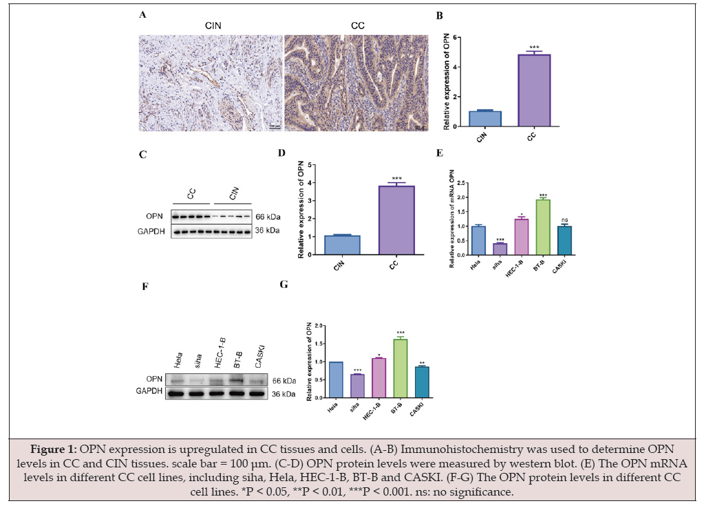
Figure 1:OPN expression is upregulated in CC tissues and cells. (A-B) Immunohistochemistry was used to determine OPN levels in CC and CIN tissues. scale bar = 100 μm. (C-D) OPN protein levels were measured by western blot. (E) The OPN mRNA levels in different CC cell lines, including siha, Hela, HEC-1-B, BT-B and CASKI. (F-G) The OPN protein levels in different CC cell lines. *P < 0.05, **P < 0.01, ***P < 0.001. ns: no significance.
EffectS of OPN on the Malignant Behaviors of CC Cells
To explore the biological functions of OPN in CC cells, siha cells were chosen for the silencing experiments of OPN, whilst BT-B cells were chosen for the overexpression experiments of OPN. As illustrated in Figure 2A-B, transfection of OE-OPN significantly elevated OPN expression, while OPN expression was markedly decreased following siRNA OPN transfection. After that, the viabilities of transfected cells were evaluated using CCK-8 assays. The results demonstrated that the overexpression of OPN remarkably facilitated cell viability (Figure 2C). The opposite results were observed when OPN was silenced (Figure 2C). As shown in Figure 3A-C, the apoptosis rate of BT-B cells with OPN overexpression were obviously reduced compared with the OE-NC group. In contrast, compared to the siRNA NC group, the apoptosis rate in the siRNA OPN group was significantly reduced. The results suggested that OPN downregulation could induce a significant promotion of CC cell apoptosis. Subsequently, we further investigated the influences of OPN on cell invasion and migration via Transwell and wound healing assays. As shown in Figure 4A-B, we found that BT-B cell migration was significantly enhanced with OPN upregulation. However, the migratory capacities were remarkably inhibited in siha cells that transfected with siRNA OPN. Furthermore, as presented in Figure 5A-D, the number of invading cells was markedly increased after OPN overexpression in BT-B cells. Conversely, OPN knockdown overtly decreased the number of invading siha cells.
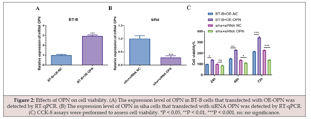
Figure 2:Effects of OPN on cell viability. (A) The expression level of OPN in BT-B cells that transfected with OE-OPN was detected by RT-qPCR. (B) The expression level of OPN in siha cells that transfected with siRNA OPN was detected by RT-qPCR. (C) CCK-8 assays were performed to assess cell viability. *P < 0.05, **P < 0.01, ***P < 0.001. ns: no significance.
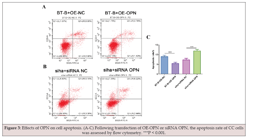
Figure 3:Effects of OPN on cell apoptosis. (A-C) Following transfection of OE-OPN or siRNA OPN, the apoptosis rate of CC cells was assessed by flow cytometry. ***P < 0.001.
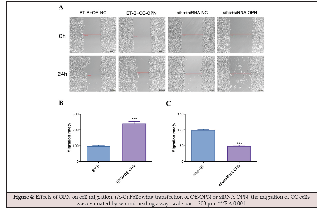
Figure 4:Effects of OPN on cell migration. (A-C) Following transfection of OE-OPN or siRNA OPN, the migration of CC cells was evaluated by wound healing assay. scale bar = 200 μm. ***P < 0.001.
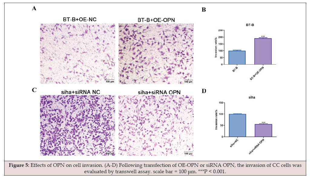
Figure 5:Effects of OPN on cell invasion. (A-D) Following transfection of OE-OPN or siRNA OPN, the invasion of CC cells was evaluated by transwell assay. scale bar = 100 μm. ***P < 0.001.
Silencing of OPN Inhibits Xenograft Growth in vivo
Consistent with the aforementioned in vitro results, knocking down OPN in nude mice remarkably inhibited the tumor growth, as evidenced by the decreased tumor volume and weight (Figure 6A-C). Afterward, HE staining was employed to observe the histopathological changes of xenografts. We found that compared to the shRNA-NC group, tumor cells in the shRNA-OPN group exhibited a reduction in size, with patchy and loose arrangement, lighter staining, and pronounced apoptosis (Figure 6D). Through immunohistochemistry, we found that injection of shRNA-OPN significantly suppressed the expression of OPN (Figure 6E-F).
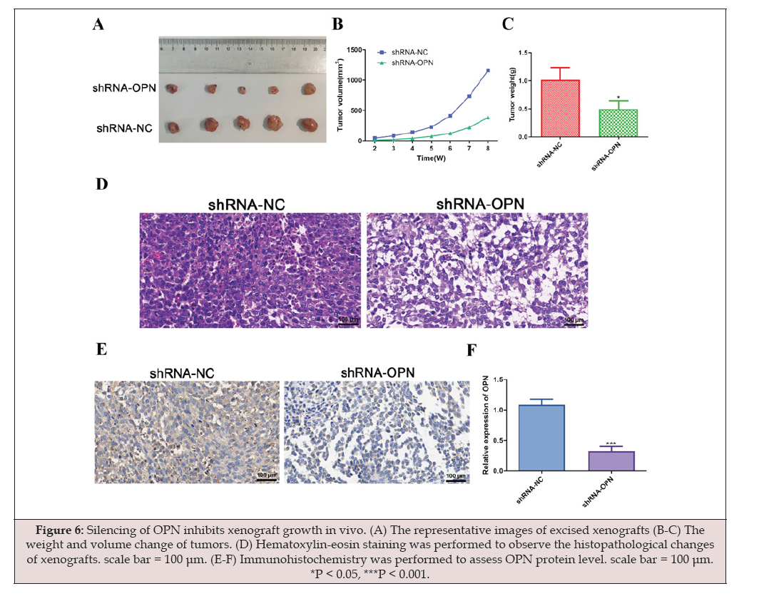
Figure 6:Silencing of OPN inhibits xenograft growth in vivo. (A) The representative images of excised xenografts (B-C) The weight and volume change of tumors. (D) Hematoxylin-eosin staining was performed to observe the histopathological changes of xenografts. scale bar = 100 μm. (E-F) Immunohistochemistry was performed to assess OPN protein level. scale bar = 100 μm. *P < 0.05, ***P < 0.001.
Discussion
OPN expression is upregulated in CC tissues and cells. (A-B) Immunohistochemistry was used to determine OPN levels in CC and CIN tissues. scale bar = 100 μm. (C-D) OPN protein levels were measured by western blot. (E) The OPN mRNA levels in different CC cell lines, including siha, Hela, HEC-1-B, BT-B and CASKI. (F-G) The OPN protein levels in different CC cell lines. *P < 0.05, **P < 0.01, ***P < 0.001. ns: no significance.
In this study, we examined the expression level of OPN in CC tissues. The results demonstrated that OPN was remarkably overexpressed in CC compared to CIN. Furthermore, to investigate the biological function of OPN in cell proliferation, apoptosis, migration and invasion, siha cells exhibiting relatively low expression levels of OPN were chosen for the following silence experiments of OPN, and BT-B cells showing relatively high expression levels of OPN were chosen for the following overexpression experiments of OPN. Significantly, limitless proliferation of tumor cells is the crucial malignant feature of cancer, which probably leads to a variation in the development of the cell cycle [20]. Huang. et al. [21] found that OPN facilitated colorectal cancer cell proliferation by activating the p38 MAPK signaling pathway. Our results identified that the upregulation of OPN by lentiviruses markedly promoted the proliferation of CC cells, and the downregulation of OPN by siRNA could suppress proliferation, as displayed by CCK-8 assay. These results suggested that OPN could promote CC cell proliferation. Additionally, the effect of OPN on the proliferation of CC was further demonstrated in CC xenograft model, which was consistent with aforementioned in vitro observations. We found that the tumors formed by siha cells transfected with OPN downregulation grew slower than the control group.
It’s widely acknowledged that the occurrence and progression of malignant tumors is involved in abnormal cell apoptosis. Decrease of OPN levels using siRNA obviously increase apoptosis in the claudin-low breast cancer cell lines [22]. Yan, et al. demonstrated that OPN can protect glioma cells from apoptosis induced by OPN siRNA through variation of the expression levels of Bcl-2 family proteins [23]. In our study, OPN was also demonstrated to have an inhibitory effect on the apoptosis of CC cells, demonstrating a significant apoptosis suppression of cancer cells following overexpression.
On the other hand, migration and invasion are crucial in cancers [24]. Migration is essential for tumor cell invasion, and the invasion of cancer cells into adjacent tissue is the first step in tumor distant metastasis. Increasing evidence suggests that OPN facilitates the migration and invasion of cancer cells during disease progression. He, et al. [25] found that the knockout of OPN in colorectal cancer remarkably inhibits cell migration. Additionally, Hao, et al. indicated that OPN activates the RON signaling pathway, thus promoting migration and invasion of non-small cell lung cancer cells [26]. In the current study, OPN expression was closely involved in the migration and invasion of CC cells. Conversely, the invasion and migration of cancer cells with OPN overexpression was significantly relieved by OPN knockdown. These data indicated that OPN can be regarded as a tumor promoter during the progression and metastasis of CC.
Conclusion
In summary, this study demonstrated that OPN can facilitate the proliferation, migration and invasion, as well as inhibit the apoptosis process in CC cells. These findings could help strengthen the theoretical foundation for creating targeted therapeutic approaches for CC in clinical practice.
Declarations
Declarations
Not applicable.
Data Availability
The datasets analyzed during the current study are available from the corresponding author on reasonable request.
Ethics Approval and Consent to Participate
The current study was approved by the ethics committee of Jinhua Maternal and Child Health Care Hospital (Approval number: 2022KY030).
Authors’ Contributions
Nengchao Xu made substantial contributions to the conception and design of the work. Huizhen Pan, Min Hu, Meixu Hu, Limei Ji, Mingxing Ding and Yu Zhang made substantial contributions to the acquisition, analysis and interpretation of data for the work. Huizhen Pan drafted the manuscript. All authors revised the manuscript critically for important intellectual content. All authors agreed to be accountable for all aspects of the work in ensuring that questions related to the accuracy and agreed final approval of the version to be published.
Funding
This work was supported by Key Social Development Projects of Science and Technology Projects in Jinhua City (2022-3-132).
Consent for Publication
Not applicable.
Conflicts of Interest
The authors declare no conflicts of interest.
References
- Sung H, Ferlay J, Siegel RL, Laversanne M, Soerjomataram I, Jemal A, et al. (2021) Global Cancer Statistics 2020: GLOBOCAN Estimates of Incidence and Mortality Worldwide for 36 Cancers in 185 Countries. CA Cancer J Clin 71(3): 209-249.
- Small W, Jr., Bacon MA, Bajaj A, Chuang LT, Fisher BJ, Harkenrider MM, et al. (2017) Cervical cancer: A global health crisis. Cancer 123(13): 2404-2412.
- Pötter R, Georg P, Dimopoulos JC, Grimm M, Berger D, Nesvacil N, et al. (2011) Clinical outcome of protocol based image (MRI) guided adaptive brachytherapy combined with 3D conformal radiotherapy with or without chemotherapy in patients with locally advanced cervical cancer. Radiother Oncol 100(1): 116-123.
- Sankaranarayanan R, Swaminathan R, Brenner H, Chen K, Chia KS, Chen JG, et al. (2010) Cancer survival in Africa, Asia, and Central America: a population-based study. Lancet Oncol 11(2): 165-173.
- Jain A, Ganesh B, Bobdey SC, Sathwara JA, Saoba S (2017) Sociodemographic and Clinical Profile of Cervical Cancer Patients Visiting in a Tertiary Care Hospital in India. Indian J Med Paediatr Oncol 38(3): 291-295.
- Rosen VM, Guerra I, McCormack M, Nogueira-Rodrigues A, Sasse A, Munk VC, et al. (2017) Systematic Review and Network Meta-Analysis of Bevacizumab Plus First-Line Topotecan-Paclitaxel or Cisplatin-Paclitaxel Versus Non-Bevacizumab-Containing Therapies in Persistent, Recurrent, or Metastatic Cervical Cancer. Int J Gynecol Cancer 27(6): 1237-1246.
- Sodek J, Ganss B, McKee MD (2000) Osteopontin. Crit Rev Oral Biol Med 11(3): 279-303.
- Chakraborty G, Jain S, Behera R, Ahmed M, Sharma P, Kumar V, et al. (2006) The multifaceted roles of osteopontin in cell signaling, tumor progression and angiogenesis. Curr Mol Med 6(8): 819-830.
- Senger DR, Asch BB, Smith BD, Perruzzi CA, Dvorak HF (1983) A secreted phosphoprotein marker for neoplastic transformation of both epithelial and fibroblastic cells. Nature 302(5910): 714-715.
- Craig AM, Bowden GT, Chambers AF, Spearman MA, Greenberg AH, Wright JA, et al. (1990) Secreted phosphoprotein mRNA is induced during multi-stage carcinogenesis in mouse skin and correlates with the metastatic potential of murine fibroblasts. Int J Cancer 46(1): 133-137.
- Rittling SR, Chen Y, Feng F, Wu Y (2002) Tumor-derived osteopontin is soluble, not matrix associated. The Journal of biological chemistry 277(11): 9175-9182.
- Suzuki K, Zhu B, Rittling SR, Denhardt DT, Goldberg HA, McCulloch CA, et al. (2002) Colocalization of intracellular osteopontin with CD44 is associated with migration, cell fusion, and resorption in osteoclasts. J Bone Miner Res 17(8): 1486-1497.
- Sharma P, Kumar S, Kundu GC (2010) Transcriptional regulation of human osteopontin promoter by histone deacetylase inhibitor, trichostatin A in cervical cancer cells. Molecular cancer 9: 178.
- Ye QH, Qin LX, Forgues M, He P, Kim JW, Peng AC, et al. (2003) Predicting hepatitis B virus-positive metastatic hepatocellular carcinomas using gene expression profiling and supervised machine learning. Nat Med 9(4): 416-423.
- Agrawal D, Chen T, Irby R, Quackenbush J, Chambers AF, Szabo M, et al. (2002) Osteopontin identified as lead marker of colon cancer progression, using pooled sample expression profiling. J Natl Cancer Inst 94(7): 513-521.
- Forootan SS, Foster CS, Aachi VR, Adamson J, Smith PH, Lin K, et al. (2006) Prognostic significance of osteopontin expression in human prostate cancer. Int J Cancer 118(9): 2255-2261.
- Tuck AB, O'Malley FP, Singhal H, Harris JF, Tonkin KS, Kerkvliet N, et al. (1998) Osteopontin expression in a group of lymph node negative breast cancer patients. Int J Cancer 79(5): 502-508.
- Singhal H, Bautista DS, Tonkin KS, O'Malley FP, Tuck AB, Chambers AF, et al. (1997) Elevated plasma osteopontin in metastatic breast cancer associated with increased tumor burden and decreased survival. Clin Cancer Res 3(4): 605-611.
- Leung DT, Lim PL, Cheung TH, Wong RR, Yim SF, Ng MH, et al. (2016) Osteopontin Fragments with Intact Thrombin-Sensitive Site Circulate in Cervical Cancer Patients. PloS one 11(8): e0160412.
- Feitelson MA, Arzumanyan A, Kulathinal RJ, Blain SW, Holcombe RF, Mahajna J, et al. (2015) Sustained proliferation in cancer: Mechanisms and novel therapeutic targets. Semin Cancer Biol 35 Suppl(Suppl): S25-s54.
- Huang RH, Quan YJ, Chen JH, Wang TF, Xu M, Ye M, et al. (2017) Osteopontin Promotes Cell Migration and Invasion, and Inhibits Apoptosis and Autophagy in Colorectal Cancer by activating the p38 MAPK Signaling Pathway. Cell Physiol Biochem 41(5): 1851-1864.
- Saleh S, Thompson DE, McConkey J, Murray P, Moorehead RA (2016) Osteopontin regulates proliferation, apoptosis, and migration of murine claudin-low mammary tumor cells. BMC Cancer 16: 359.
- Yan W, Qian C, Zhao P, Zhang J, Shi L, Qian J, et al. (2010) Expression pattern of osteopontin splice variants and its functions on cell apoptosis and invasion in glioma cells. Neuro Oncol 12(8): 765-775.
- Novikov NM, Zolotaryova SY, Gautreau AM, Denisov EV (2021) Mutational drivers of cancer cell migration and invasion. Br J Cancer 124(1): 102-114.
- He B, Mirza M, Weber GF (2006) An osteopontin splice variant induces anchorage independence in human breast cancer cells. Oncogene 25(15): 2192-2202.
- Hao C, Cui Y, Chang S, Huang J, Birkin E, Hu M, et al. (2019) OPN promotes the aggressiveness of non-small-cell lung cancer cells through the activation of the RON tyrosine kinase. Scientific reports 9(1): 18101.




 We use cookies to ensure you get the best experience on our website.