
Mini Review 
 Creative Commons, CC-BY
Emerging Roles of Hypoxia-Inducible Factor and Glucose Metabolism in Esophageal Cancer: Possible Targets of Cancer
*Corresponding author:Mingxing Ding, Medical Molecular Biology Laboratory, School of Medicine, Jinhua University of Vocational Technology, 1188 Wuzhou Road, Jinhua, Zhejiang, P.R. China.
Received:March 10, 2026; Published:March 17, 2026
DOI: 10.34297/AJBSR.2026.30.003944
Abstract
Hypoxia-Inducible Factors (HIFs) are important nuclear transcription factors that are activated in solid tumors, such as esophageal cancer. HIF plays a central role in regulating energy metabolism. Currently, the cause of esophageal cancer remains unclear; it may be related to eating coarse food, drinking, smoking, genetics, and esophagitis. Because cancer cells are regulated by multiple survival pathways, research on these regulatory pathways can uncover potential cancer targets. During rapid tumor growth, the insufficient blood supply leads to a hypoxic tumor environment. Many studies have shown that cancer has two major characteristics: HIF-1 expression is upregulated, and metabolic pathways are altered. Even with sufficient oxygen, malignant tumor cells prefer glycolysis to oxidative phosphorylation. This metabolic characteristic of aerobic glycolysis is called the ‘Warburg Effect’. Malignant tumor cells exhibit high glucose uptake, active glycolysis, and high lactic acid production. The nearly universal characteristic of primary and metastatic cancers is the upregulation of glycolysis, which results in increased glucose consumption to meet the demands of tumor cells for energy, biosynthesis, and redox signaling. Therefore, in this article, we review recent research on HIF and glucose metabolism in esophageal cancer, which may provide targets for new treatment strategies to improve the therapeutic outcome.
Keywords: Hypoxia, Esophageal cancer, HIF, Glucose metabolism, Warburg effect, Metastasis
Abbreviations:ESCC: esophageal squamous cell carcinoma; RT-qPCR: real-time quantitative polymerase chain reaction; IHC: Immuno histo chemistry; HIF-1α: hypoxia-inducible factor-1 alpha; HIF-2α: hypoxia-inducible factor-2 alpha; GLUT-1: glucose transporter-1; HK-Ⅱ: hexokinase-Ⅱ; LDHA: lactate dehydrogenase-A; Epo: erythropoietin; Epo-R: erythropoietin receptor; VEGF: vascular endothelial growth factor.
Introduction
Esophageal Cancer (EC) is the ninth most common cancer worldwide [1]. Patients with esophageal cancer, whether Esophageal Squamous Cell Carcinoma (ESCC) or Esophageal Adenocarcinoma (EAC), have a relatively high mortality rate [2] that ranks sixth among all cancers, and their five-year survival rate is only 15%–25% [3]. Early detection of esophageal cancer is difficult, and patients are usually diagnosed at an advanced stage [4]. Highly proliferating tumor cells, such as esophageal tumor cells, require a large energy supply [5]. Anaerobic conditions in the tumor can decrease the amount of ATP produced, thus decreasing energy production. Therefore, in order to adapt to this energy shortage, metabolic pathways are modified to let tumor cells upregulate glycolysis. Hypoxia-Inducible Factor (HIF) signaling and metabolic pathway modification can facilitate multiple processes: rapid tumor cell proliferation, neovascularization, invasion and metastasis, and evasion of apoptosis [6]. Traditional treatment options for esophageal cancer include metastatic chemotherapy. However, approximately half of the patients receiving chemoradiation therapy have local recurrence. In order for radiation therapy to be effective, molecular oxygen is essential. Under normoxic conditions, ionizing radiation generates free radicals and Reactive Oxygen Species (ROS) that damage DNA [7].
Malignant tumor cells are distinct from normal cells of the body because they have the ability to self-proliferate and resist apoptosis. They also have unlimited replication potential, are insensitive to anti-growth signals, and can form a dense vascular network. They can invade, metastasize, and perform aerobic glycolysis. HIF-1 is a core transcription factor that regulates tumor cell adaptation to hypoxia and participates in the transcriptional regulation of many genes during it [8]. Among the major characteristics of tumors, carrying out aerobic glycolysis is critical in giving them great survival advantages.
Basic Structure of HIF
HIF is a heterodimeric protein composed of two subunits, HIF-α and HIF-β, and is the main protein that regulates the response of cells to changes in oxygen levels. The HIF- α family includes: HIF-1α, HIF-2α, and HIF-3α, that are stabilized by oxygen. Under hypoxic conditions, these three isoforms can heterodimerize with HIF-1β to modulate the transcription of target genes [9]. The center of the HIF-1α subunit contains an Oxygen-Dependent Degradation Domain (ODDD); its C-terminus is a transactivation site, which includes two Transactivation Domains (TADs), TAD-N (amino acids 531-575) and TAD-C (amino acids 786-826). The region between the two TAD sequences is an inhibitory domain (ID, amino acids 576-785), which inhibits the transcriptional activation of TAD [10]. Although the regulatory mechanism of HIF-1α synthesis is not dependent on O2, its degradation is. Under normoxic conditions, HIF-1α is rapidly degraded by the intracellular oxygen-dependent ubiquitin protease, and under hypoxic conditions, its expression is induced and maintained [11]. The oxygen-dependent and oxygen- independent regulation of HIF-1α can lead to angiogenesis, metastasis, and cell survival [12,13].
The Role of HIF in Tumors
Hypoxia is an important biological condition that surrounds solid tumor cells. Under hypoxic conditions, gene transcription and expression undergo significant changes in the tumor cells, to adapt to the environment. These genes are the transcriptional targets of HIF-1α. Previous research has found that HIF-1α has more than 70 target genes that are involved in the growth of various types of tumors. The target genes regulated by HIF-1α that promote tumor growth participate in glucose metabolism, cell proliferation, angiogenesis, cell invasion, and other biological behaviors [14].
HIF, Glucose Transport, and Glycolysis
Changes in the energy metabolism of tumors play a prominent role in malignant transformation. Compared to the aerobic oxidation of normal cells, tumor cells under normoxic or hypoxic conditions preferentially use glycolysis to meet metabolic demands [15]. Based on this characteristic, Positron Emission Tomography (PET) technology is used to diagnose malignant tumors in clinical practice. It is widely accepted that HIF-1 is an important mediator in the adaptive response to hypoxia, and is capable of activating and regulating a variety of downstream genes. Among them, genes responsible for regulating energy metabolism include glucose transporters 1 (GLUT1) and 3 (GLUT3), prolyl-4-hydroxylase α1, phosphofructokinase L, lactate dehydrogenase A, aldolases A and C, pyruvate kinase M, enolase 1, hexokinase 1 and 2, and Glyceraldehyde Phosphate Dehydrogenase (GAPDH) [16,17]. Hexokinase is the first enzyme in the glycolysis pathway and irreversibly converts glucose to glucose-6-phosphate, thereby allowing glucose molecules to enter the glycolytic cycle. HIF-1 mainly plays a role in glycolysis [18]. By binding to the target gene, HIF-1 induces the gene expression of glycolytic enzymes, promotes anaerobic metabolism, and facilitates energy production through glycolysis in tumor cells [19]. Thus, current research on the effects of HIF-1 on energy metabolism mainly focuses on tumor cells.
HIF, Tumor Cell Proliferation, and Apoptosis
Studies have shown that an inhibitor of HIF-1α, PX-478, can significantly inhibit proliferation and promote apoptosis of ESCC cells, thus reducing tumor volume and showing significant antitumor activity [20]. On the contrary, HIF-1α-mediated activation of lnc191 transcription promotes the growth and metastasis of ESCC in vitro and in vivo [21]. Another study showed that, in addition to regulating glucose metabolism of tumor cells, HIF-1 can also upregulate multiple growth factors such as erythropoietin, insulin-like growth factor 2, transforming growth factor α, and cyclin D1, allowing liver cancer cells to proliferate, differentiate, and adapt to the hypoxic microenvironment [22].
HIF and Vascularization
The rapid rate of tumor growth leads to a lack of oxygen due to an imbalance between oxygen consumption and insufficient vasculature; thus, tumor hypoxia develops in solid tumors. In many malignant tumors, Vascular Endothelial Growth Factor (VEGF) is one of the main drivers of angiogenesis and is a major factor in the development and maintenance of vascular networks [23]. VEGF increases oxygen supply by directly increasing vascular permeability and inducing the formation of new blood vessels [24]. Studies have shown that there is a positive correlation between HIF-1α and VEGF expression in esophageal tumors, and the expression of HIF-1α and VEGF is significantly upregulated in tumor cells under hypoxia [25]. In an esophageal Three-Dimensional (3D) cell culture system, HIF-1α plays an important role in tumor adaptation and tolerance to the hypoxic microenvironment by directly regulating its target gene VEGF [26].
HIF And Tumor Invasion and Metastasis
Tumor invasion and metastasis are highly dependent on Matrix Metalloproteinases (MMPs) [27]. Under hypoxic conditions, hypoxia-induced upregulation of MMPs greatly enhances the invasive capacity of tumor cells, and causes ECM stiffness and degradation [28]. Shao et al [25] demonstrated that HIF-1α and VEGF play a synergistic role in the invasion and metastasis, and consequent malignancy of esophageal cancer, thus reducing the survival rate of patients with oral squamous cell carcinoma.
HIF And Tumor Resistance to Radiotherapy
Radiation therapy is the standard of care for Esophageal Squamous Cell Carcinoma (ESCC), especially in patients with inoperable tumors in the upper and middle esophagus. Several studies have confirmed that cancer cells may be 2-3 times more sensitive to radiation under normoxic conditions than under hypoxic conditions [29,30]. Many preclinical and clinical studies have shown that tumor cells can be protected from radiotherapy by VEGF [31]. A study of the combination of bortezomib with radiotherapy for esophageal cancer showed that it effectively sensitized ESCC cells to radiation by reducing the expression of HIF-1α and VEGF, activating caspase to induce apoptosis, and delaying DNA damage repair [32]. Similarly, Brucea Javanica Oil Emulsion (BJOE) can inhibit the expression of HIF-1α, alleviate hypoxia, and increase the sensitivity of ESCC to radiotherapy [33].
HIF and the Prognosis of Esophageal Cancer
Esophageal squamous cell carcinoma is a solid tumor, and its internal hypoxia often leads to increased HIF-1α protein expression, which is related to the invasion and metastasis of esophageal cancer [34]. High HIF-1α expression is correlated with poor prognosis in patients with ESCC, suggesting its association with an increased risk of esophageal squamous cell carcinoma. Furthermore, HIF-1α expression is a potential indicator of lymph node metastasis and correlates with tumor stage. HIF-1 is a key transcription factor that transmits hypoxic signals and mediates the effects of hypoxia [35]. HIF-1α regulates both autocrine or paracrine signaling pathways for cell growth to promote angiogenesis in hypoxic conditions [36]. HIF-1α is a functional subunit of HIF-1, it participates in the transcriptional regulation of various target genes and affects the energy metabolism, proliferation, and apoptosis of tumor cells [15,37]. Downstream HIF-1 signaling generates a series of reactions in cells, enabling it to adapt to the hypoxic environment, and promoting tumor vascularization [38]. It also increases tumor invasiveness and resistance to radiotherapy and chemotherapy and participates in tumor progression [39,40]. Unfortunately, the role of HIF-1α in the invasiveness and prognosis of esophageal cancer is still unclear, and research results vary among different cancers, ethnicities, and populations. To date, most clinical studies have relied on immunohistochemical methods and have found that the positive expression rate of HIF-1 is relatively high, between 43.75% and 69.9% [25,41,42]. The positive expression rate is significantly higher in squamous cell carcinoma than in adenocarcinoma [43]. The positive expression rate of HIF-1α is also higher than that of HIF-2α [44]. Increasing research has shown that the positive expression of HIF-1α protein in ESCC is higher in patients at stage T3 or T4 with lymph node metastasis than in patients at stage T1 or T2 without lymph node metastasis [25,45]. Furthermore, HIF- 1α protein expression increases with TNM staging [46]. Univariate analysis identified high positive HIF-1α expression as a risk factor for tumor invasion, lymph node and distant metastasis, lymphatic invasion, and positive surgical margins. Cox regression multivariate analysis showed that HIF-1α expression was an independent prognostic factor for 5-year survival. However, opposing results have also been reported [25]. Munipalle et al [43] used immunohistochemistry and found varying levels of HIF-1α expression in 36 patients with ESCC. Furthermore, regression analysis showed that HIF-1α and was HIF-2α not an independent prognostic factor for survival [47,48]. Although the sample size was small, this study indicated that HIF-1α may not have prognostic value in European populations of patients with ESCC. Additionally, in 53 patients with esophageal cancer who received esophagectomy, although HIF-1 mRNA expression was related to protein expression and upregulated to varying degrees in esophageal squamous and adenocarcinoma cells, it was not correlated with histomorphology or prognosis, and the expression did not predict tumor progression, remission, or prognosis [49]. Therefore, the complicated relationships between hypoxia-related molecules HIF-1α, HIF-2α, GLUT-1, RAC-1, and BECLIN- 2 and angiogenesis factors VEGF, VEGF-C, VEGF-D, CD34, and E-cadherin in esophageal cancer are still under research [25,42,49- 51]. Additional clinical evidence is needed to show clinical utility in the early diagnosis, treatment, and prognosis of esophageal cancer.
HIF Inhibitors and Esophageal Cancer (Treatment)
Semenza and Wang first discovered Hypoxia-Inducible Factor (HIF)-1, established the structure, and determined the coding sequence of its cDNA, which provided the foundation for developing HIF-1 inhibitors against tumors [52]. According to the mechanism of action, HIF inhibitors can be divided into regulators of: HIF-1α mRNA expression, HIF-1α protein translation, HIF-1α protein degradation, HIF-1α DNA binding, and HIF-1α transcriptional activity. PX-478 (S-2-amino-3- [4’-N, N, -bis(chloroethyl)amino] phenyl propionic acid N-oxide dihydrochloride) is a selective inhibitor that inhibits the transcription and translation of HIF-1α. PX-478 inhibits tumor growth in vivo and in vitro, induces cell cycle arrest at the G2 phase, promotes apoptosis, and reduces expression of COX-2 and PD-L1 in ESCC cells [20]. In recent years, the discovery and development of new small molecules targeting HIF-1α have been an exciting direction in the development of therapy, and relevant studies have increased exponentially [12, 53]. However, the main challenge in developing small-molecule HIF-1α inhibitors is their specificity. Hence, further research is needed to explore the potential of HIF-1α inhibitors in esophageal cancer.
Conclusion
Even though living standards have improved, factors such as poor dietary habits and polluted living environments continue to contribute to an increase in the number of patients with esophageal cancer. Esophageal cancer remains a significant cause of cancer-related deaths worldwide. In some Western countries, its incidence has increased dramatically, with a 5-year survival rate of only 10% to 15% [54]. Most patients are already in an advanced stage at the time of diagnosis and cannot be treated with radical surgical resection. Early-stage esophageal cancer can be removed with surgery. In its middle and advanced stages, where the cancer has already metastasized, resection complemented with neoadjuvant chemotherapy is the main choice of treatment. High-dose chemotherapy drugs can kill cancer cells and reduce tumor volume. Acquired resistance to radiation remains a major obstacle in increasing the survival rate of Esophageal Cancer (EC) [55]. Hypoxia-related factors play a crucial role in resistance to radiation, and 2ME2 inhibits the expression of HIF-1α in ECA-109 cells, sensitizing them to radiation [56]. These findings provide an important foundation for probing into the biological function of HIF as a potential therapeutic target for the treatment of esophageal cancer. Tumor cell metabolism differs from that of normal cells. For example, under hypoxic conditions, HIF-1α can inhibit Pyruvate Dehydrogenase (PDH) by activating Pyruvate Dehydrogenase Kinase (PDK), leading to an accumulation of pyruvate in the cytoplasm [57]. At the same time, the expression of LDH induced by HIF-1α can promote the metabolic cycle of glycolytic cofactor NAD+ by catalyzing the transformation of pyruvate to lactic acid, thereby promoting continuous glycolysis and the production of ATP [15,58-61].
Changes in the glucose metabolism of tumors play a significant role in tumor progression and HIF is one of the key enzymes in glycolysis. The evaluation of hypoxia in esophageal cancer is important for predicting treatment outcomes and efficacy. Many studies have demonstrated that HIF was associated with differentiation and lymph node metastasis, and with the TNM stage, HIF could be a potential independent prognostic factor (Table 1). Novel strategies for the therapeutic targets of HIF in esophageal cancer are to be further explored and may play a role in improving treatment outcomes.
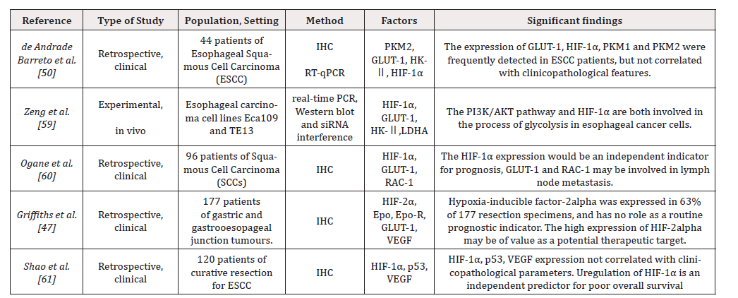
Table 1:Hypoxia inducible factor and Warburg effect in esophageal carcinoma.
Table Abbreviations: ESCC: esophageal squamous cell carcinoma, RT-qPCR: real-time quantitative polymerase chain reaction IHC: Immunohistochemistry, HIF-1α: hypoxia-inducible factor-1 alpha, HIF-2α: hypoxia-inducible factor-2 alpha, GLUT-1: glucose transporter-1, HK-Ⅱ: hexokinase-Ⅱ, LDHA: lactate dehydrogenase-A, Epo: erythropoietin, Epo-R: erythropoietin receptor, VEGF: vascular endothelial growth factor
Acknowledgements
This work was supported by the Science and Technology Project of Jinhua, China (grant no. 2026-3-015).
Conflict of Interest
The authors declare no conflicts of interest
References
- Sheikh M, Roshandel G, McCormack V, Malekzadeh R (2023) Current Status and Future Prospects for Esophageal Cancer. Cancers (Basel) 15(3): 765.
- Fatehi Hassanabad A, Chehade R, Breadner D, Raphael J (2020) Esophageal carcinoma: Towards targeted therapies. Cell Oncol (Dordr) 43(2): 195-209.
- Lin EW, Karakasheva TA, Hicks PD, Bass AJ, Rustgi AK (2016) The tumor microenvironment in esophageal cancer. Oncogene 35(41): 5337-5349.
- Deboever N, Jones CM, Yamashita K, Ajani JA, Hofstetter WL (2024) Advances in diagnosis and management of cancer of the esophagus. BMJ 385: e074962.
- Shafaee A, Dastyar DZ, Islamian JP, Hatamian M (2015) Inhibition of tumor energy pathways for targeted esophagus cancer therapy. Metabolism 64(10): 1193-1198.
- Li Y, Zhang Y, Li X, Huang Y, Chen W, et al. (2021) Status of hypoxia-inducible factor-1α expression in non-small cell lung cancer. Pharmazie 76(9): 404-411.
- Srinivas US, Tan BWQ, Vellayappan BA, Jeyasekharan AD (2019) ROS and the DNA damage response in cancer. Redox biology 25: 101084.
- Balamurugan K (2016) HIF-1 at the crossroads of hypoxia, inflammation, and cancer. International journal of cancer 138(5): 1058-1066.
- Masson N, Ratcliffe PJ (2003) HIF prolyl and asparaginyl hydroxylases in the biological response to intracellular O (2) levels. Journal of cell science 116 (Pt 15): 3041-3049.
- Jiang BH, Zheng JZ, Leung SW, Roe R, Semenza GL (1997) Transactivation and inhibitory domains of hypoxia-inducible factor 1alpha. Modulation of transcriptional activity by oxygen tension. The Journal of biological chemistry 272(31): 19253-19260.
- Kim J, So D, Shin HW, Chun YS, Park JW (2015) HIF-1 α Upregulation due to Depletion of the Free Ubiquitin Pool. Journal of Korean medical science 30(10): 1388-1395.
- Masoud GN, Li W (2015) HIF-1 α pathway: role, regulation and intervention for cancer therapy. Acta pharmaceutica Sinica B 5(5): 378-389.
- Rashid M, Zadeh LR, Baradaran B, Molavi O, Ghesmati Z, et al. (2021) Up-down regulation of HIF-1α in cancer progression. Gene 798: 145796.
- Semenza GL (2012) Hypoxia-inducible factors: mediators of cancer progression and targets for cancer therapy. Trends in pharmacological sciences 33(4): 207-214.
- Singh D, Arora R, Kaur P, Singh B, Mannan R, et al. (2017) Overexpression of hypoxia-inducible factor and metabolic pathways: possible targets of cancer. Cell Biosci 7: 62.
- Haigis MC, Deng CX, Finley LW, Kim HS, Gius D (2012) SIRT3 is a mitochondrial tumor suppressor: a scientific tale that connects aberrant cellular ROS, the Warburg effect, and carcinogenesis. Cancer research 72(10): 2468-2472.
- Semenza GL (2010) HIF-1: upstream and downstream of cancer metabolism. Current opinion in genetics & development 20(1): 51-56.
- Yu L, Lu M, Jia D, Ma J, Ben Jacob E, et al. (2017) Modeling the Genetic Regulation of Cancer Metabolism: Interplay between Glycolysis and Oxidative Phosphorylation. Cancer research 77(7): 1564-1574.
- Hochwald JS, Zhang J (2017) Glucose Oncometabolism of Esophageal Cancer. Anti-cancer agents in medicinal chemistry 17(3): 385-394.
- Zhu Y, Zang Y, Zhao F, Li Z, Zhang J, et al. (2017) Inhibition of HIF-1α by PX-478 suppresses tumor growth of esophageal squamous cell cancer in vitro and in vivo. American journal of cancer research 7(5): 1198-1212.
- Wei S, Fan X, Li X, Zhou W, Zhihua Zhang, et al. (2025) Hypoxia Induced Lnc191 Upregulation Dictates the Progression of Esophageal Squamous Cell Carcinoma by Activating GRP78/ERK Pathway. Adv Sci (Weinh) 12(4): e2406674.
- Xu Z, Liu E, Peng C, Li Y, He Z, et al. (2012) Role of hypoxia-inducible-1α in hepatocellular carcinoma cells using a Tet-on inducible system to regulate its expression in vitro. Oncol Rep 27(2): 573-578.
- Patel SA, Nilsson MB, Le X, Cascone T, Jain RK, et al. (2023) Molecular Mechanisms and Future Implications of VEGF/VEGFR in Cancer Therapy. Clin Cancer Res 29(1): 30-39.
- Zhu H, Feng Y, Zhang J, Zhou X, Hao B, et al. (2011) Inhibition of hypoxia inducible factor 1α expression suppresses the progression of esophageal squamous cell carcinoma. Cancer Biol Ther 11(11): 981-987.
- Shao JB, Li Z, Zhang N, Yang F, Gao W, et al. (2019) Hypoxia-inducible factor 1α in combination with vascular endothelial growth factor could predict the prognosis of postoperative patients with oesophageal squamous cell cancer. Pol J Pathol 70(2): 84-90.
- Terashima J, Sampei S, Iidzuka M, Ohsakama A, Tachikawa C, et al. (2016) VEGF expression is regulated by HIF-1α and ARNT in 3D KYSE-70, esophageal cancer cell spheroids. Cell Biol Int 40(11): 1187-1194.
- Niland S, Riscanevo AX, Eble JA (2021) Matrix Metalloproteinases Shape the Tumor Microenvironment in Cancer Progression. Int J Mol Sci 23(1): 146.
- Najafi M, Farhood B, Mortezaee K (2019) Extracellular matrix (ECM) stiffness and degradation as cancer drivers. J Cell Biochem 120(3): 2782-2790.
- Sorensen BS, Busk M, Olthof N, Speel EJ, Horsman MR, et al. (2013) Radiosensitivity and effect of hypoxia in HPV positive head and neck cancer cells. Radiother Oncol 108(3): 500-505.
- Xie Y, Zhang J, Ye S, He M, Ren R, et al. (2012) SirT1 regulates radiosensitivity of hepatoma cells differently under normoxic and hypoxic conditions. Cancer Sci 103(7): 1238-1244.
- Barker HE, Paget JT, Khan AA, Harrington KJ (2015) The tumour microenvironment after radiotherapy: mechanisms of resistance and recurrence. Nat Rev Cancer 15(7): 409-425.
- Wang D, Qin Q, Jiang QJ, Wang DF (2016) Bortezomib sensitizes esophageal squamous cancer cells to radiotherapy by suppressing the expression of HIF-1α and apoptosis proteins. J Xray Sci Technol 24(4): 639-646.
- Pan P, Yang BX, Ge XL (2018) Brucea javanica seed oil enhances the radiosensitivity of esophageal cancer by inhibiting hypoxia-inducible factor 1α, in vitro and in vivo. Oncology letters 15(3): 3870-3875.
- Abdel Wahab AF, Mahmoud W, Al Harizy RM (2019) Targeting glucose metabolism to suppress cancer progression: prospective of anti-glycolytic cancer therapy. Pharmacol Res 150: 104511.
- Hayashi Y, Yokota A, Harada H, Huang G (2019) Hypoxia/pseudohypoxia-mediated activation of hypoxia-inducible factor-1α in cancer. Cancer Sci 110(5): 1510-1517.
- Tsai HJ, Wilson JE (1996) Functional organization of mammalian hexokinases: both N- and C-terminal halves of the rat type II isozyme possess catalytic sites. Archives of biochemistry and biophysics 329(1): 17-23.
- Tang NN, Zhu H, Zhang HJ, Zhang WF, Jin HL, et al. (2014) HIF-1α induces VE-cadherin expression and modulates vasculogenic mimicry in esophageal carcinoma cells. World J Gastroenterol 20(47): 17894-17904.
- Manuelli V, Pecorari C, Filomeni G, Zito E (2022) Regulation of redox signaling in HIF-1-dependent tumor angiogenesis. FEBS J 289(18): 5413-5425.
- Kimura S, Kitadai Y, Tanaka S, Kuwai T, Hihara J, et al.(2004) Expression of hypoxia-inducible factor (HIF)-1alpha is associated with vascular endothelial growth factor expression and tumour angiogenesis in human oesophageal squamous cell carcinoma. Eur J Cancer 40(12): 1904-1912.
- Wigerup C, Pahlman S, Bexell D (2016) Therapeutic targeting of hypoxia and hypoxia-inducible factors in cancer. Pharmacol Ther 164: 152-169.
- Guo X, Chen Y, Fang W, Yang W, Shi L, et al. (2013) Metastasis associated protein 1 correlates with Hypoxia inducible-factor 1 alpha expression and lymphangiogenesis in esophageal cancer. Thoracic cancer 4(3): 312-317.
- Chai DM, Bao ZQ, Hu JG, Ma L, Feng ZZ, et al. (2013) Vasculogenic mimicry and aberrant expression of HIF-lα/E-cad are associated with worse prognosis of esophageal squamous cell carcinoma. J Huazhong Univ Sci Technolog Med Sci 33(3): 385-391.
- Munipalle PC, Viswanath YK, Davis PA, Scoones D (2011) Prognostic value of hypoxia inducible factor 1α in esophageal squamous cell carcinoma. Dis Esophagus 24(3): 177-181.
- Koukourakis MI, Giatromanolaki A, Skarlatos J, Corti L, Blandamura S, et al. (2001) Hypoxia inducible factor (HIF-1a and HIF-2a) expression in early esophageal cancer and response to photodynamic therapy and radiotherapy. Cancer research 61(5): 1830-1832.
- Kurokawa T, Miyamoto M, Kato K, Cho Y, Kawarada Y, et al. (2003) Overexpression of hypoxia-inducible-factor 1alpha (HIF-1alpha) in oesophageal squamous cell carcinoma correlates with lymph node metastasis and pathologic stage. Br J Cancer 89 (6): 1042-1047.
- Ogane N, Yasuda M, Shimizu M, Miyazawa M, Kamoshida S, et al. (2010) Clinicopathological implications of expressions of hypoxia-related molecules in esophageal superficial squamous cell carcinoma. Ann Diagn Pathol 14(1): 23-29.
- Griffiths EA, Pritchard SA, McGrath SM, Valentine HR, Price PM, et al. (2008) Hypoxia-associated markers in gastric carcinogenesis and HIF-2alpha in gastric and gastro-oesophageal cancer prognosis. Br J Cancer 98(5): 965-973.
- Matsuyama T, Nakanishi K, Hayashi T, Yoshizumi Y, Aiko S, et al. (2005) Expression of hypoxia-inducible factor-1alpha in esophageal squamous cell carcinoma. Cancer Sci 96(3): 176-182.
- Chen Y, Lu Y, Lu C, Zhang L. (2009) Beclin-1 expression is a predictor of clinical outcome in patients with esophageal squamous cell carcinoma and correlated to hypoxia-inducible factor (HIF)-1alpha expression. Pathol Oncol Res 15(3): 487-493.
- de Andrade Barreto E, de Souza Santos PT, Bergmann A, de Oliveira IM, Wernersbach Pinto L, et al. (2016) Alterations in glucose metabolism proteins responsible for the Warburg effect in esophageal squamous cell carcinoma. Exp Mol Pathol 101(1): 66-73.
- Zhou X, Xia Q, Chen M, Zhang X, Huang M, et al. (2024) THBS1 promotes angiogenesis and accelerates ESCC malignant progression by the HIF-1/VEGF signaling pathway. Cell Biol Int 48(3): 311-324.
- Wang GL, Jiang BH, Rue EA, Semenza GL (1995) Hypoxia-inducible factor 1 is a basic-helix-loop-helix-PAS heterodimer regulated by cellular O2 Proceedings of the National Academy of Sciences of the United States of America 92(12): 5510-5514.
- Rashid M, Zadeh LR, Baradaran B, Molavi O, Ghesmati Z, et al. (2021) Up-down regulation of HIF-1α in cancer progression. Gene 798: 145796.
- Moyana TN, Janoski M (1996) Recent trends in the epidemiology of esophageal cancer. Comparison of epidermoid- and adenocarcinomas. Annals of clinical and laboratory science 26(6): 480-486.
- Malhotra A, Sharma U, Puhan S, Chandra Bandari N, Anjali Kharb, et al. (2019) Stabilization of miRNAs in esophageal cancer contributes to radioresistance and limits efficacy of therapy. Biochimie 156: 148-157.
- Lu YR, Song J, Zhabihula BX, Zhang JR (2019) 2-Methoxyestradiol promotes radiosensitivity of esophageal squamous cell carcinoma by suppressing hypoxia-inducible factor-1α European review for medical and pharmacological sciences 23(24): 10785-10795.
- Kim JW, Tchernyshyov I, Semenza GL, Dang CV (2006) HIF-1-mediated expression of pyruvate dehydrogenase kinase: a metabolic switch required for cellular adaptation to hypoxia. Cell metabolism 3(3): 177-185.
- Wang M, Chen H, He X, Zhao X, Zhang H, et al. (2022) Artemisinin inhibits the development of esophageal cancer by targeting HIF-1α to reduce glycolysis levels. J Gastrointest Oncol 13(5): 2144-2153.
- Zeng L, Zhou HY, Tang NN, Zhang WF, He GJ, et al. (2016) Wortmannin influences hypoxia-inducible factor-1 alpha expression and glycolysis in esophageal carcinoma cells. World J Gastroenterol 22(20): 4868-4880.
- Ogane N, Yasuda M, Shimizu M, Miyazawa M, Kamoshida S, et al. (2010) Clinicopathological implications of expressions of hypoxia-related molecules in esophageal superficial squamous cell carcinoma. Ann Diagn Pathol 14(1): 23-29.
- Shao N, Han Y, Song L, Song W (2020) Clinical significance of hypoxia-inducible factor 1α, and its correlation with p53 and vascular endothelial growth factor expression in resectable esophageal squamous cell carcinoma. J Cancer Res Ther 16(2): 269-275.




 We use cookies to ensure you get the best experience on our website.