
Research Article 
 Creative Commons, CC-BY
Does the Timing of Intrauterine Fetal Irradiation Affect the Child’s Subsequent General Somatic Morbidity?
*Corresponding author:Alexander N Stojarov, Department of Radiation Medicine and Ecology, Belarusian State Medical University, 220116, Minsk, Belarus, Tel: +375297560223.
Received:March 20, 2026; Published:March 25, 2026
DOI: 10.34297/AJBSR.2026.30.003952
Abstract
The incidence of morbidity in children exposed in utero due to incorporation of radioactive iodine by their pregnant mothers alters subsequent morbidity depending on the timing of radiation exposure. The highest thyroid absorbed doses occur in children during the first and third trimesters of pregnancy. In fetuses, the highest doses occur during the second trimester. The predominant morbidity types in children are respiratory and endocrine disorders. The highest morbidity in females was recorded if intrauterine irradiation occurred during the second trimester of pregnancy, and in males, during the third trimester. Differences in the prevalence of certain morbidity classes were also observed. These findings can be explained by the influence of I-131 on gene expression, with subsequent changes in endocrine system activity involving thyroid hormones.
Keywords: Ionizing radiation, I-131, Morbidity, Thyroid gland, Pregnancy, Embryogenesis
Abbreviations:TG: Thyroid Gland; TSH: Thyroid Stimulating Hormone.
Introduction
As it is known, the radiation disaster at the Chernobyl nuclear power plant was accompanied by the release of uranium nuclear decay products into the environment, including iodine isotopes, primarily I-131. This isotope, which enters the human body through inhalation, as well as with food, mainly with whole milk, accumulates in the thyroid gland (TG), which leads to its irradiation. Previously, using the example of a cohort of women living in the Stolin district of the Brest region, who at various stages of pregnancy in late April 1986, i.e., as a result of the accident at the Chernobyl nuclear power plant, were exposed to a radioactive cloud containing iodine isotopes, we studied their morbidity over the next 30 years [1]. It was found that after a fairly long period of time, compared with a control group of women who were pregnant after the decay of I-131, the incidence of cardiovascular pathology and respiratory pathology increases [2]. Cardiovascular pathology was predominant, manifested by primary hypertension [3]. Both essential hypertension and respiratory diseases were characterized by a dependence on the absorbed thyroid dose. Embryogenesis is known to be characterized by specific stages of development. The potential for a particular effect from intrauterine irradiation depends on the nature of the ionizing radiation, the absorbed dose, and the stage of fetal development at the time of irradiation [4]. The stage of intrauterine development is particularly important because differentiation of systems and organs occurs at specific times, and this will determine the type of damage to the fetus and, consequently, subsequent morbidity.
The analysis of the health status of children born by irradiated women is of a particular interest, depending on the timing of their exposure to radiation during intrauterine development.
We were able to discover the relationship between women irradiated as a result of the Chernobyl accident and their children over three decades. Accordingly, the aim of the present study was to analyze the incidence of different pathologies in individuals irradiated being fetuses at different stages of intrauterine development in comparison with a cohort of individuals who were born later, when I-131 had almost completely decayed.
Materials and Methods
The main group of individuals was comprised of residents of the Stolin District of the Brest Region, born by women exposed to a radioactive cloud that passed through this region of Belarus in late April-early May 1986. The cloud contained iodine radionuclides, including I-131. The main cohort included 123 individuals, including 62 females and 61 males. Their dates of birth ranged from June 3, 1986, to February 6, 1987. The average absorbed dose to the thyroid gland in the overall group was 32.2 ± 3.31, with a median of 22 mGy; for males, it was 35.4 ± 5.4, with a median of 23 mGy; for females, it was 29.1 ± 3.9, with a median of 22 mGy. In this study, the comparison group also included residents of the Stolin District of the Brest Region, but those who have been born later. It included 121 individuals from the same district, who were identical to the experimental group not only in terms of residence but also in social status. These individuals included 57 males and 56 females. Their dates of birth ranged from January 3, 1988, to December 31, 1988.
Verified health data for irradiated and non-irradiated individuals were obtained from the State Registry of Persons Affected by the Chernobyl Accident. Only primary morbidity was considered in the study. The International Classification of Diseases, 10th Revision (ICD-10) was used.
Statistical data processing was performed using the software applications Statistika 10.0 (StatSoft.Inc, USA) and SigmaPlot 12.5 (Systat Software Inc., Germany).
Results and Discussion
Our primary interest was the analysis of the distribution of thyroid gland absorbed doses at different stages of pregnancy in women and their children. Figure 1 presents these data. As it can be seen, absorbed doses in mothers ranged from 75-85 mGy, while in fetuses, doses ranged from 10-45 mGy. The maximum doses to the fetus occurred in the second trimester of pregnancy.
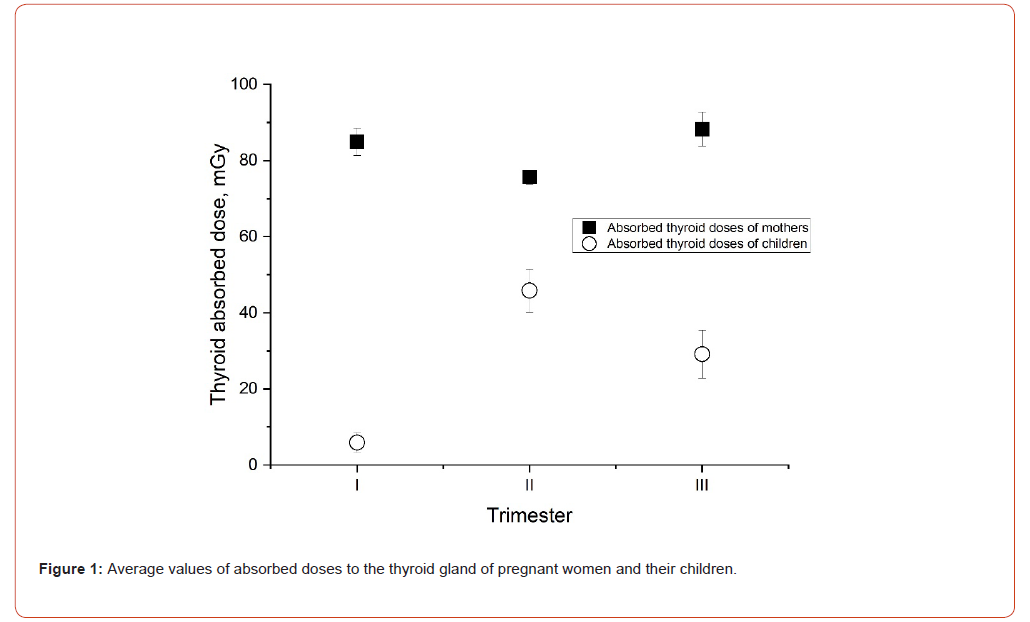
Figure 1:Average values of absorbed doses to the thyroid gland of pregnant women and their children.
The distribution of absorbed doses in mothers by trimester showed that the highest doses, ranging from 71-85 mGy, occurred in first and second trimesters of pregnancy (Figure 2). However, in the most unfavorable period, i.e., the first trimester of pregnancy, due to the beginning of organ development, thyroid doses to the fetus were minimal, which is a positive factor for potential longterm consequences.
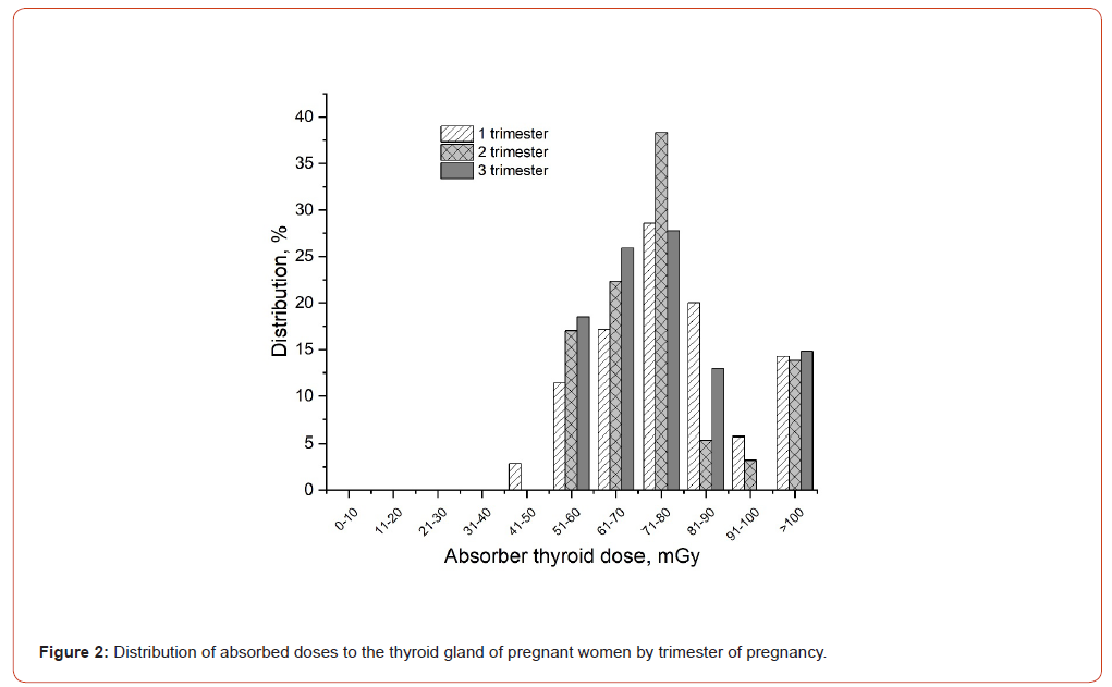
Figure 2:Distribution of absorbed doses to the thyroid gland of pregnant women by trimester of pregnancy
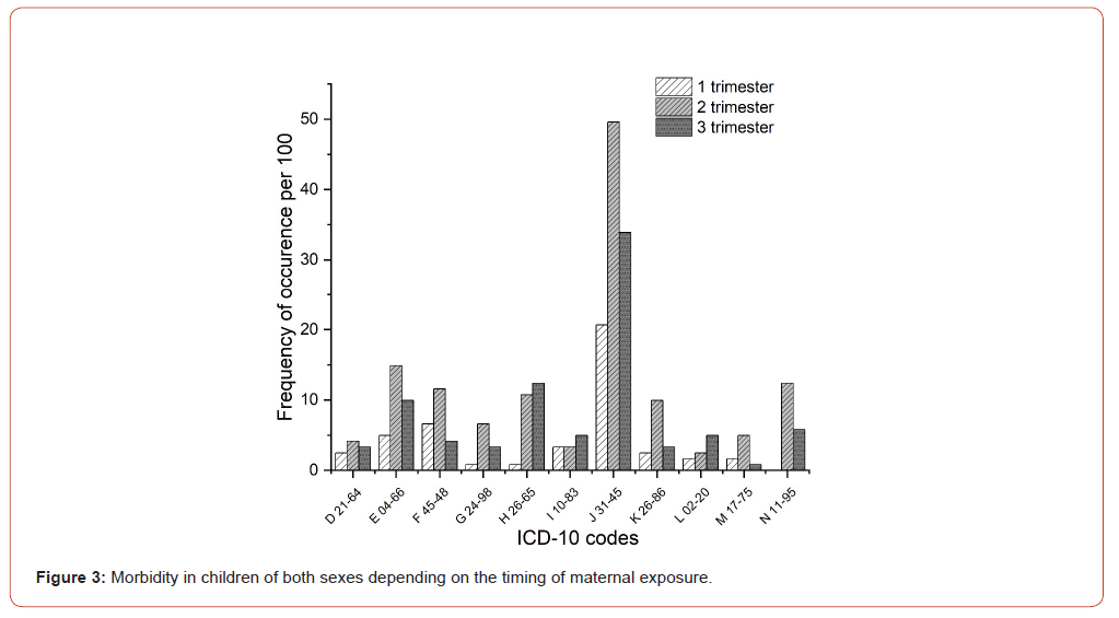
Figure 3:Morbidity in children of both sexes depending on the timing of maternal exposure.
An analysis of the incidence of diseases in children of both sexes irradiated in utero revealed that the highest incidence of diseases, particularly respiratory diseases, was recorded in children exposed to radioactive iodine in the second trimester of pregnancy (Figure 3). This pathology was somewhat less common in children exposed to radioactive iodine in the third trimester of pregnancy. Endocrine and genitourinary diseases were the second and third most common. It should be noted that, as with the aforementioned types of pathology, a predominance of diseases in other classes (mental and behavioral disorders, diseases of the nervous system, digestive system, musculoskeletal system, and bone tissue) was observed if fetal exposure also occurred in the second trimester of pregnancy.
It was also quite interesting to perform the analysis of morbidity dependence on the gender of the fetus irradiated in utero. Figure 4 show these data.
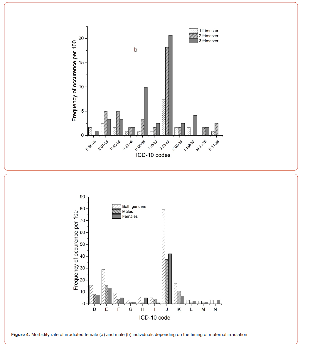
Figure 4:Morbidity rate of irradiated female (a) and male (b) individuals depending on the timing of maternal irradiation.
It is clearly evident that the dominant period causing increased morbidity among irradiated females is the second trimester of pregnancy. This applies to the same types of pathologies observed in the mixed-gender group. In contrast, among males, the dominant period causing increased morbidity was most often the third trimester of pregnancy. This applied to most classes of diseases, with the exception of endocrine and genitourinary diseases, as well as mental and behavioral disorders. Among females irradiated in utero, respiratory and endocrine pathologies predominated, while among males, respiratory diseases and diseases of the eye and adnexa were more common. They also had a higher incidence of skin and subcutaneous tissue diseases compared to females.
In such an analysis, it is necessary to consider the incidence of diseases in individuals not exposed to radioactive iodine. These individuals, as noted above, were individuals of similar social status but born after the radioactive iodine had decayed. Figure 5 presents these data. It is clear that the predominant pathologies in unexposed individuals over the long term are also respiratory and endocrine diseases. Moreover, for some pathologies, the incidence rate even exceeds that one of irradiated children. This concerns pathologies of the endocrine, respiratory, and gastrointestinal systems.
Explanations for these findings are necessary. The increased incidence of respiratory diseases in both irradiated and nonirradiated individuals is fairly easily explained. This pathology was recorded in all groups throughout the post-Chernobyl years, but was most common in 1991-1997, when children were 5 to 11 years old. As it is well known, this pathology is most common in childhood, especially in children attending group institutions such as kindergartens and schools.
An analysis of the morbidity pattern in irradiated children revealed that the predominant pathology was associated with hypertrophy of the tonsils and adenoids (J35.3). Acute respiratory diseases—acute bronchitis and acute pharyngitis—ranked second and third. This order of pathology types differed from that of children born later and not exposed to radioactive iodine (I-131). In these individuals, acute respiratory diseases (acute bronchitis J20.9, acute nasopharyngitis J00.9, and acute pharyngitis J02.9) ranked first.
Regarding the increased incidence in the group of nonirradiated children, the following can be hypothesized. In our opinion, two body systems may be involved in this phenomenon. Firstly, the immune system, which, under the influence of radiation, can block the immediate immune response to infectious agents. Secondly, the endocrine system of the body may have played a certain role, the center of damage of which was the thyroid gland, which had incorporated radioactive iodine. Previously, during a study of the effects of I-131 in thyroid cells, 27 reversibly regulated genes were discovered [5]. A number of genes (Pax8, Sic5a5, Tg, Tpo), playing an important role in the functioning of this organ, the synthesis of its hormones and the influence on the metabolism of peripheral cells, have low expression activity at low doses formed by I-131 and change their activity with increasing radiation exposure. It is known that during different trimesters of pregnancy, many biochemical parameters change, including the levels of T4, T3, thyroid-stimulating hormone (TSH), etc. Thus, at the end of the first trimester of pregnancy (at 10 weeks), the TSH level drops to minimum values, then, as the pregnancy period increases (by 20 weeks), its level returns to normal. Accordingly, the level of T4 in the woman’s body will also change. Against the background of a decrease in the TSH level, an insufficient level of transmembrane iodide transfer in thyrocytes will negatively affect the state of the thyroid system of the woman and the fetus, synthesis of hormones and, due to the known instability of the genome, can cause changes in the function of peripheral organs, including immune cells, lymphoid tissue, affecting inflammatory processes, oxidative stress, triggering the process of cell regeneration, which can cause the observed hypertrophy of this tissue [6].
Conclusion
Thus, thyroid irradiation during fetal development, depending on the timing of exposure to radioactive iodine, affects subsequent morbidity in the individual differently. In females, the highest incidence of morbidity in the late period is associated with irradiation during the second trimester of pregnancy. In males, increased morbidity is associated with irradiation during the third trimester of maternal pregnancy. The highest thyroid doses in mothers occurred in the first and second trimesters of pregnancy, while the lowest absorbed doses in their fetuses occurred in the first trimester. The predominant morbidity in all cases was respiratory and endocrine disorders. These findings can be explained by the influence of I-131 on gene expression, with subsequent changes in endocrine system functioning involving thyroid hormones.
Conflict of Interest
Authors have no conflicts of interest.
Funding
This study did not receive any special grant from any fundig agency in the government, commercial or non-profit sector.
References
- Stojarov AN, Khrustalev VV, Odinzova Stojarova DA (2021) Non-Cancer Morbidity of Women Irradiated During Pregnancy Due to the Incorporation of I-131 as a Result of the Chernobyl Accident. Intl J Clin Stud & Med Case Reps 10: 1-3.
- Stojarov AN, Khrustalev VV (2022) Delayed Consequences of The Radioactive Iodine Intake by Pregnant Women from The Side of Respiratory System. AJBSR 15 (2): 210-216.
- Stojarov AN, Khrustalev VV, Adzintsova Stazharava DA (2021) Cardiovascular Pathology in Women Who Received Radioactive Iodine Exposure During Pregnancy as a Result of the Chernobyl Accident. Biomed J Sci & Tech Res 37(2): 29320-29328.
- Dekaban AS (1968) Abnormalities in children exposed to x-radiation during various states of gestation: tentative time table of radiation injury in the human fetus. J Nucl Med Part 1 9: 471-477.
- Rudqvist N (2015) Radiobiological effects of the thyroid gland. University of Gothenburg, Gothenburg. pp.69.
- Jara EL, Muñoz Durango N, Llanos C, Carlos Fardella, Pablo A González, et al. (2017) Modulating the function of the immune system by thyroid hormones and thyrotropin. Immun Lett 184: 76-83.




 We use cookies to ensure you get the best experience on our website.