
Review Article 
 Creative Commons, CC-BY
3D Guided Screw Placement in the Spine and Pelvis – Who Bears Responsibility? – A User’s Perspective
*Corresponding author:Bandar Khalid Alrais, BSc. Biochemistry, master’s in forensic and toxicology, PhD in health management, Saudi Arabia.
Received:August 18, 2025; Published:August 22, 2025
DOI: 10.34297/AJBSR.2025.28.003661
Abstract
Thyrotoxicosis is an illness diagnosed with clinically in decreased amount of Thyroid Stimulating Hormone (TSH) and increased serum Triiodothyronine (T3) and Thyroxine (T4) levels [1,2]. Low or high TSH concentrations and low cognitive performance have been associated [3] Hyperthyroid causes increased resting energy consumption, loss of weight, decreased cholesterol levels, increased lipolysis, and development of hypermetabolic conditions characterized by gluconeogenesis [2,3]. These studies have revealed that all concept of thyrotoxicosis.
Introduction
The thyroid gland, located in the anterior portion of the neck just below and bilateral to the thyroid cartilage, develops from the thyroglossal duct and portions of the ultimobranchial body [4-6]. The right lobe of thyroid gland is normally larger than the left [7] and, in some individuals, a superior portion of glandular tissue, or pyramidal lobe, can be identified. Thyroid tissue may be found anywhere along the path of the thyroglossal duct, from its origin on midline posterior portion of the tongue to its termination of thyroid gland in the neck [4,6] The thyroglossal duct passes through the region of the developing hyoid bone, and remnants of the duct can become enclosed or surrounded by the bone [7] Ectopic thyroid tissue may secrete thyroid hormones or become cystic or neoplastic [8] In a few individuals, the only functional thyroid tissue is in these ectopic locations [5] (Figure 1).
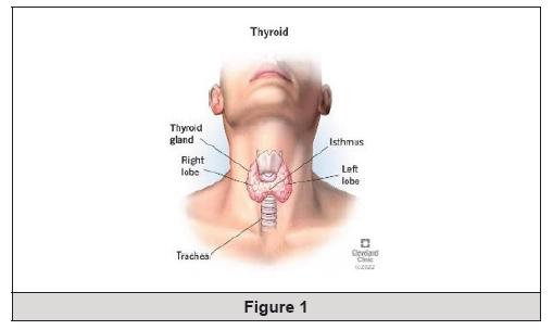
Figure 1:
Thyrotoxicosis is a common disease worldwide. The incidence rate of thyrotoxicosis is influenced by a number of factors, of which the iodine intake in the population may be important. In areas with increased iodine intake level [9,10], hypothyroidism is more common than hyperthyroidism, whereas hyperthyroidism dominates in areas with mild and moderate iodine deficiency [3]. Thyrotoxicosis is divided into a number of oncological subtypes with different etiology, clinical presentation, prognosis and outcome of therapy [3,9].
Thyrotoxicosis is characterized by high thyroid hormone synthesis and secretion from the thyroid gland, whereas thyrotoxicosis refers to the clinical syndrome of excess circulating thyroid hormones, irrespective of the source. Thyrotoxicosis showed low serum TSH level and high serum levels of T4 or T3, or both [10].
Thyrotoxicosis is two subcategories: thyrotoxicosis with hyperthyroidism and thyrotoxicosis without hyperthyroidism. Thyrotoxicosis with hyperthyroidism is named Graves’ disease and toxic nodular goiter (Plummer’s disease), and is characterized by a high thyroid radioactive iodine uptake. Other, thyrotoxicosis without hyperthyroidism happened by painless and sub-acute thyroiditis, iodine- induced and drug-induced thyroid dysfunction, and factitious ingestion of excess thyroid hormones [11,12].
Diagnosis of thyroid diseases is often influenced by various manifestations that are non-specific such as lethargy, weight gain or loss, palpitations or weakness. A factor applied in the diagnosis of thyroid diseases such as hypothyroidism or hyperthyroidism is concentration level interpretation of thyroid hormone T4, T3 and TSH that are commonly measured in a test called Thyroid Function Test (TFT) [10]. In a subgroup of patients, the interpretation of TFT is more accurate, either because the results appear to not correlate with the clinical picture i.e. a low TSH in patient with hypothyroid or normal TFT in patients is suspected to be strongly thyrotoxicosis. In these patients, a structured approach that includes clinical reassessment of thyroid status, along with various potentiating factors [e.g. infection, stress, drug therapy] can help in identifying the possible solution to this discordant TFT [11,13].
Thyrotoxicosis occurs due to an inappropriately high synthesis and secretion of Thyroid Hormone (TH) by the thyroid [13]. TH increases tissue thermogenesis and the basal metabolic rate and reduces serum cholesterol levels and systemic vascular resistance. The complications of thyrotoxicosis include weight loss, osteoporosis, fragility fractures, atrial fibrillation, embolic events, and cardiovascular dysfunction [14] (Figure 2).
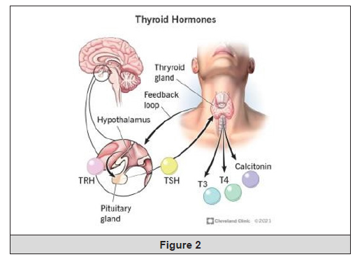
Figure 2:
Thyrotoxicosis is the term applied when there is excess thyroid hormone in the circulation due to any reason. The diagnosis of hyperthyroidism is generally straightforward, with raised serum thyroid hormones T3, T4 and low serum TSH in almost all cases [14,15]. Thyrotoxicosis can be easily diagnosed by a high serum level of T4 and T3 and a low serum level of TSH [15]. Hyperthyroidism is confirmed by a high isotope (I 131 or Tc99) uptake by the thyroid gland, while in thyroiditis it will be low [16].
Appropriate treatment of hyperthyroidism occur by identification of the cause. Anti-thyroid drugs, radioactive iodine, and surgery are the traditional treatments for the 3 common forms of hyperthyroidism. Beta-adrenergic blocking agents are used in most patients for symptomatic relief and might be the only treatment needed for thyroiditis, which is transient [16,17].
Symptoms of hyperthyroidism which caused by thyrotoxicosis heat intolerance, palpitations, anxiety, fatigue, weight loss, muscle weakness, and, in women, irregular menses. Clinical findings may include tremor, tachycardia, lid lag, and warm moist skin. Symptoms and signs of subclinical hyperthyroidism, if present, are usually vague and nonspecific [18,19,20].
Type of Thyrotoxicosis
Thyrotoxicosis is defined as the state of thyroid hormone elevated and is not synonymous with hyperthyroidism, which is the result of high thyroid hormones. However, the major etiologies of thyrotoxicosis are hyperthyroidism caused by Graves’ disease, toxic multi-nodular goiter, and toxic adenomas. To treat thyrotoxicosis appropriately, define the cause is essential [21,22].
Grave’s Disease
Graves’ disease is an autoimmune disorder in which Thyroid Stimulating Immunoglobulin (TSI) binds to and stimulates the TSH receptor on the thyroid cell membrane, resulting in excessive synthesis and secretion of thyroid hormone [23]. Graves’ disease is frequently complicated by ophthalmopathy, which can be a debilitating component of the disease, resulting in impaired quality of life [23,24]. The management of Graves’ disease treated restore the patient to a eu- thyroid state and minimize the extent of extra-thyroidal manifestations such as ophthalmopathy (Figure 3).
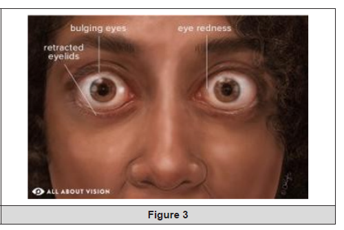
Figure 3:
Patients with Graves’ disease usually have diffuse, nontender, symmetrical enlargement of the thyroid gland. Ophthalmopathy, include protrusion of the eyes with periorbital soft tissue swelling and inflammation, and inflammatory changes in the extraocular muscles resulting in diplopia and muscle imbalance, is clinically evident in 30% of patients with Graves’ disease [25]. Graves’ disease is the most common cause of hyperthyroidism in developed countries. It is an autoimmune condition in which antibodies developed against the TSH receptor which is reason for unopposed stimulation of the thyroid gland [25,26]. Circulating thyroid antibodies activate TSH receptor and stimulate thyroid follicular hypertrophy and hyperplasia lead to high thyroid hormone production [23,27]. Graves’ disease cause hyperthyroidism and diffuse goiter, ophthalmopathy, pretibial myxedema and thyroid acropachy may also be observed [25]. The pathogenesis of this enigmatic condition remains incompletely understood but the central pathogenetic event is the unregulated stimulation of the TSH receptor by autoreactive TSH Receptor Antibodies (TRAbs). Graves’ disease showed predominantly affects women (female: male ratio 8:1), typically in their 3rd to 5th decades of life [23, 25]. People with a family history of hyperthyroidism or other autoimmune diseases such as pernicious anemia, myasthenia gravis, type I diabetes mellitus, and celiac disease have an increased propensity of developing Graves [28] (Figure 4).
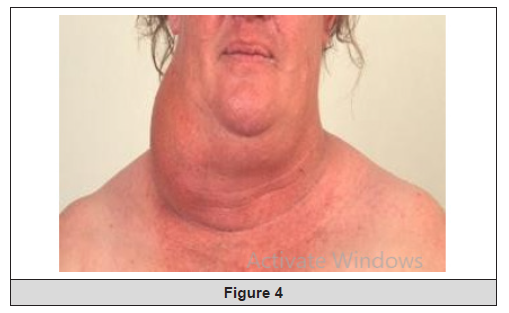
Figure 4:
Pathogenesis of Grave’s Disease: The cause of Graves’ disease remains unclear, but it is believed to result from a complex interaction between genetic background (heredity), environmental factors and the immune system [29,30]. Without cause, the immune system produces an antibody (TSH Receptor Antibody (TRAb) that stimulates the thyroid gland to manufacture excess thyroid hormone. Genetic susceptibility to the disease is thought to be polygenic [30]. Graves’ disease has been reported to be associated with the Human Leukocyte Antigen (HLA) gene on chromosome 6p, the Cytotoxic T Lymphocyte Antigen-4 (CTLA-4) gene on chromosome 2q33, and the lymphoid Tyrosine Phosphatase (PTPN22) gene on chromosome 1p13. In Graves’ disease, hyperthyroidism results from the action of Thyroid-Stimulating Antibodies (TSAb) directed against the thyrotropin receptor on the surface of the thyroid cell. The Thyroid-Stimulating Immunoglobulin (TSI) binds to and stimulates the TSH receptor on the thyroid cell membrane resulting in follicular cell growth, vascularity increase, and in excessive synthesis and secretion of thyroid hormone. The thyroid gland typically displays lymphocytic infiltration, with T- lymphocyte abnormality and absence of follicular destruction. T cells activate local inflammation and tissue remodeling by producing and releasing cytokines, leading to B-cell dysregulation and increase in autoantibody production. An imbalance between pathogenic and regulatory T cells is thought to be involved in both the development of Graves’ disease and its severity [30,31].
Toxic Multinodular Goiter (TMNG)
Toxic Multinodular Goiter (TMNG) is an important cause of hyperthyroidism. It is caused by unwarranted release of thyroid hormones from multiple autonomously functioning nodules in the thyroid gland. It is more common in areas of dietary iodine deficiency (third-world countries) and in the elderly (poor diet). TMNG is more common than Graves’ disease in the elderly [23].
Toxic Nodular Goitre (toxic adenoma)
Toxic nodular goitre is the most frequent cause of thyrotoxicosis in the elderly, especially in iodine deficient areas. Solitary toxic nodules are more common in women than in men with 1:5 M: F ratio reported in some studies. An autonomous thyroid nodule (toxic adenoma) is a discrete thyroid mass whose function is independent of pituitary control [23, 24].
Subacute Thyroiditis
Thyroiditis is characterized by a self-limiting course of thyrotoxicosis, followed by hypothyroidism, and then returns to normal thyroid function or is inflammation of the thyroid gland that typically follows a viral upper respiratory infection and causes additional release of preformed thyroid hormone. It is slightly more common in females than males (ratio of 1.5:1) and permanent hypothyroidism occurs in 10-20% of cases. Acute painful thyroiditis often presents following a respiratory tract infection, while painless thyroiditis may occur post- partum in up to 9% of otherwise normal women [23-25].
Suppurative Thyroiditis
Suppurative thyroiditis is an infection of the thyroid gland typically caused by bacteria but can be caused by fungus, mycobacteria, or parasites. It is most common in immunocompromised persons or those with underlying thyroid disease. It presents with a tender erythematous anterior neck mass, fever, dysphagia, and dysphonia [11,23].
Drug Induced Hyperthyroidism
Amiodarone-Induced Hyperthyroidism (AIH): Amiodarone, a benzofuranic derivative containing 75 mg of iodine per 200-mg tablet, is widely used for the long-term treatment of cardiac arrhythmias. Amiodarone inhibits the 5’monodeiodination of T4 in the liver and pituitary, thereby decreasing serum T3 and mildly increasing serum T4 levels without altering TSH concentrations. Type 1 AIT is due to the high iodine content of amiodarone. It occurs in areas of iodine deficiency and in patients with underlying thyroid disorders, such as multinodular goiter. In Type 1 AIT, the thyroid gland produces and releases excessive amounts of thyroid hormone. In contrast, Type 2 AIT results from a destructive process in the thyroid gland in which preformed thyroid hormones leak from the damaged follicular cells in patients without underlying thyroid disease. Amiodarone induced thyrotoxicosis is more common in iodine deficient areas and appears to be more common in men [11,23].
Amiodarone-Induced Hyperthyroidism (AIH) is much more common and challenging to treat than amiodarone-induced hypothyroidism. Amiodaroneinduced thyrotoxicosis results from two different mechanisms. The iodine released during the metabolism of the drug is responsible for the thyrotoxicosis in most cases. Predisposing factors include micronodular and macronodular goiter, which are common in older patients who most often require amiodarone. Thyroid autoimmunity has also been incriminated as a predisposing factor and antithyroid antibodies have been found following amiodarone administration in some patients but not in others [23].
Other drugs that cause thyrotoxicosis include interferon-α, lithium, tyrosine kinase inhibitors, highly active antiretroviral therapies, immune checkpoint mediators and the humanized monoclonal antibodies used in the treatment of multiple sclerosis. Although these drugs may cause transient thyrotoxicosis through destructive thyroiditis the immune modifying agents such as interferon-α, HAART, and alemtuzumab may in addition induce Graves’ diseases through less well-defined immune reactivation mechanisms [1,2].
Postpartum Thyroiditis
Postpartum thyroiditis is inflammation of the thyroid gland following delivery. It is a transient form of hyperthyroidism that can develop 6 weeks to 6 months postpartum with a significant chance of recurrence in subsequent pregnancies. Patients are present with painless goiter and typically have significant family history of autoimmune disease [29].
Diagnosis
Direct tests of thyroid function involve the adminis tration of radioactive iodine [3,10]. Measurement of the thyroid Radioactive Iodine Uptake (RAIU) is the most common of these tests. 131I has been used for this test, but 123I is preferred because it exposes the patient to a lower radiation dose. The RAIU is measured 24 hours after the administration of the isotope. The RAIU varies inversely with the plasma iodide concentration and directly with the functional state of the thyroid. In the United States the normal 24-hour RAIU is 10% to 30%. The RAIU discriminates poorly between normal and hypothyroid states. Values above the normal range use ally indicate thyroid hyperfunction.6 The measurement of the basal serum TSH concern tration is useful in the diagnosis of hyperthyroidism and hypothyroidism [3,10] Very sensitive methods are now available, such as immunoradiometric or hemalum nascent, to measure serum TSH. In cases of hyperthy roidism the TSH level is almost always low or none testable. Higher levels indicate hypothyroidism, and lower levels signify hyperthyroidism [1]. Three types of thyroid autoantibodies can be mean surged for diagnostic uses. The autoantibodies that can be tested for are Thyroglobulin (Tg Ab), Peroxidase (TPO Ab), and TSH Receptor (TSHR Ab). 2 A thyroid scan is a common test used to localize thyroid nodules and to locate functional ectopic thyroid tissue. 123Ior 99Tc is injected, and a scanner localizes areas of radioactive concentration. This technique al lows for the identification of nodules 1cm or larger [9]. Several tests are available that measure the thyroid hormone concentration and binding in blood. Highly specific and sensitive radioimmunoassay are used to measure serum T4 and T3 concentrations and rarely to measure Reverse Triiodothyronine (rT3) concentration [3,10] Elevated levels usually indicate hyperthyroid ism, and lower levels usually indicate hypothyroidism. The free hormone levels usually correlate better with the metabolic state than do total hormone levels. Indi rect assays are used to estimate the free T4 level.6 Current practice is to screen patients suspected of being hyperthyroid with the TSH serum level and measure or estimate the free T4 concentration [12,13]. A decreased TSH level and an increased free T4 concentration are classic for hyperthyroidism [12]. Some patients are hyperthyroid with a low TSH level and normal free T4 concentration, but they have an elevated free T3 level. A few patients have a normal or elevated TSH and a high free T4. These patients either have a TSH-secreting pituitary adenoma or have thyroid hormone– resistance syndrome [3, 12,13].
Treatment / Management
The recommended treatment of thyrotoxicosis depends on its underlying cause. Beta- blockers, such as propranolol, are used to alleviate adrenergic symptoms such as sweating, anxiety, and tachycardia. Theoretically, propranolol inhibits 5′-monodeiodinase, thus blocking the peripheral conversion of T4 to T3. Propranolol also blocks both β1- and β2 receptors, making it a first-line agent. However, it should generally be avoided in patients with asthma [18,22].
The 3 mainstays of treatment include thionamide drugs, radioiodine therapy, and thyroid surgery.
Thionamide Drugs
Thionamide drugs, including Propylthiouracil (PTU) and methimazole, cause decreased the production of thyroid hormone by acting as preferential substrates for thyroid peroxidase. At high doses, PTU also inhibits the peripheral conversion of T4 to T3 [18,22].
Methimazole is prescribe at a dosage of 15 to 30mg/d for 4 to 8 weeks in the treatment of Graves disease, during which most patients achieve a euthyroid state. Once euthyroid is established, treatment can proceed with 1 of 2 approaches. In the block-replace method, the same dose of thioamide is continued to inhibit thyroid hormone production, while levothyroxine is added to maintain euthyroid. Alternatively, the thioamide dose can be gradually reduced to allow endogenous thyroid hormone synthesis, thereby sustaining a euthyroid state [22, 23].
In Graves disease, long-term remission is achieved in about 50% of patients treated with thioamide drugs. However, a drawback of thioamides is the uncertainty of relapse after discontinuation. Prolonged treatment beyond 18 months has not been shown to improve remission rates. Methimazole is more effective and has a longer half-life than PTU, allowing for once-daily dosing [26].
Additionally, PTU carries a higher risk of hepatotoxicity. Agranulocytosis occurs in approximately 1 in 300 patients treated with thioamides and typically presents with symptoms such as sore throat, mouth ulcers, and high fever. A differential white blood cell count is recommended for all patients on thioamides experiencing febrile illness or pharyngitis. Minor adverse effects of thioamides include pruritus, arthralgia, and gastrointestinal upset. If patients continue to have anti-TSH receptor antibodies or signs of hyperthyroidism after 18 months of treatment, options such as radioiodine therapy or surgery should be considered [23,26].
Teprotumumab, an IGF1R blocker, is approved by the Food and Drug Administration (FDA) for the treatment of Graves orbitopathy. Teprotumumab also shows potential for treating Graves disease and other forms of thyrotoxicosis [23].
Radioiodine Therapy
Radioiodine therapy is the most common treatment for adults with Graves disease in the United States. This therapy is also effective for treating follicular nodules and toxic multinodular goiter [18,23,26]. Radioactive iodine is administered as a single oral dose. This is absorbed by the thyroid gland, causing tissue-specific inflammation that leads to thyroid fibrosis and a gradual destruction of thyroid tissue over the following months. Hypothyroidism typically develops within 6 to 12 months, need lifelong levothyroxine therapy for most patients. Patients with large goiters, severe thyrotoxicosis, ischemic heart disease, heart failure, or arrhythmia are advised to undergo thioamide pretreatment until achieving a euthyroid state before receiving radioiodine therapy [18,23].
Notably, radiation therapy is contraindicated during pregnancy and lactation, and conception should be avoided for 6 to 12 months for both males and females. A small risk of thyrotoxicosis exacerbation exists in the first month after treatment due to the release of preformed thyroid hormones. Additionally, radioiodine therapy is a definitive risk factor for developing Graves orbitopathy [23,25].
The treatment of thyroiditis differs because antithyroid drugs are ineffective, as patients typically have reduced production of new thyroid hormones. Thyroiditis is usually transient, and the treatment is focused on symptom control using beta-blockers. In cases of subacute thyroiditis, Nonsteroidal Anti-Inflammatory Drugs (NSAIDs) and, occasionally, systemic glucocorticoids may be prescribed to manage pain and inflammation. Beta-blockers are recommended for older patients with symptomatic thyrotoxicosis and also for any thyrotoxic patient with a resting heart rate exceeding 90 bpm or with underlying cardiovascular disease [3,10].
Children with thyrotoxicosis may be treated with methimazole, radioiodine therapy, or thyroidectomy. Methimazole is the first-line therapy for Graves disease in children, typically administered for 1 to 2 years, as some children may achieve remission. Radioiodine therapy is not recommended for children aged 5 or younger. Additionally, PTU should be avoided in children due to its risk of hepatotoxicity [3,23].
Thyroid Surgery
Thyroidectomy, whether total or partial, provides rapid and effective treatment for thyrotoxicosis. However, it is invasive and expensive and also results in permanent hypothyroidism, necessitating lifelong levothyroxine therapy. Pretreatment to achieve euthyroid is recommended before surgery to minimize the risk of exacerbating thyrotoxicosis or triggering a thyroid storm. Surgery is indicated for cases of hyperthyroidism resistant to medical therapy, significant thyroid enlargement causing compressive symptoms, or when thyroid cancer is suspected. The surgical procedure usually involves either a total thyroidectomy, where the entire thyroid gland is removed, or a thyroid lobectomy, which involves removing one lobe of the thyroid (right or left). Most patients can resume normal activities within a few days. Common complications include transient hypocalcemia due to temporary hypoparathyroidism and vocal cord paresis caused by injury to the recurrent laryngeal nerve [3,10,23].
Acknowledgements
None.
Conflict of Interest
None.
References
- Adams R, Oh ES, Yasar S, Lyketsos CG, Mammen JS (2023) Endogenous and Exogenous Thyrotoxicosis and Risk of Incident Cognitive Disorders in Older Adults. JAMA Intern Med 183(12): 1324-1331.
- Smith TJ, Hegedüs L (2016) Graves' Disease. N Engl J Med 375(16): 1552- 1565.
- Sharma A, Stan MN (2019) Thyrotoxicosis: Diagnosis and Management. Mayo Clin Proc 94(6): 1048-1064.
- Little JW (2002) Thyroid disease. In: Little JW, Falace DA, Miller CS, Rhodus NL, editors. Dental management of the medically com promised patient. 6th St Louis: Mosby; pp: 283- 303.
- Larsen PR, Davies TF, Schlumberger MJ (2003) Thyroid physiology and diagnostic evaluation of patients with thyroid disorders. In: Larsen PR, Kronenberg HM, Melmed D, Polonsky KS, editors. Williams textbook of endocrinology. 10th Philadelphia: W. B. Saunders; pp: 331-65.
- Jameson JL, Weetman AP (2005) Disorders of the thyroid gland: anat omy and development. In: Kasper DL, Braunwald E, Fauci AS, Hauser SL, Longo DL, Jameson JL, et al. editors. Harrison’s principles of internal medicine. 16th New York: McGraw Hill; pp: 2105, Chapter 320.
- Mazzaferri EL (1986) The thyroid. In: Mazzaferri EL, editor. Endocri nology. 3rd New York: Medical Examination Publishing.
- Shin K, Kim J, Park J, Oh TJ, Kong SH, et al. (2023) A machine learning-assisted system to predict thyrotoxicosis using patients' heart rate monitoring data: a retrospective cohort study. Sci Rep 13(1): 21096.
- Smith TJ, Hegedüs L (2017) Graves' Disease. N Engl J Med 376(2): 185.
- Kravets I (2016) Hyperthyroidism: Diagnosis and Treatment. Am Fam Physician 93(5): 363-370.
- Campennì A, Avram AM, Verburg FA, Iakovou I, Hänscheid H, et al. (2023) The EANM guideline on radioiodine therapy of benign thyroid disease. Eur J Nucl Med Mol Imaging 50(11): 3324-3348.
- Razvi S, Bhana S, Mrabeti S (2019) Challenges in Interpreting Thyroid Stimulating Hormone Results in the Diagnosis of Thyroid Dysfunction. J Thyroid Res 2019: 4106816.
- Jung CK, Bychkov A, Kakudo K (2022) Update from the 2022 World Health Organization Classification of Thyroid Tumors: A Standardized Diagnostic Approach. Endocrinol Metab (Seoul) 37(5): 703-718.
- Beck-Peccoz P, Giavoli C, Lania A (2019) A 2019 update on TSH-secreting pituitary adenomas. J Endocrinol Invest 42(12): 1401-1406.
- Beck-Peccoz P, Persani L, Lania A, Feingold KR, Ahmed SF, Anawalt B, et al. (2022) Thyrotropin-Secreting Pituitary Adenomas. In: Endotext [Internet]. South Dartmouth (MA): MDText.com, Inc.; Oct 13, 2022.
- Chivukula KK, Toro-Tobón D, Motazedi B, Goyal R (2021) Thyroid storm as an early presentation of hCG-producing metastatic choriocarcinoma: a case report and review of the literature. BMJ Case Rep 14(9): 242868.
- Anfinsen OG, Lima K (2021) Amiodarone-induced thyrotoxicosis. Tidsskr Nor Laegeforen 141(16).
- Bahn RS, Burch HB, Cooper DS, Garber JR, Greenlee MC, et al. (2011) American Thyroid Association. American Association of Clinical Endocrinologists. Hyperthyroidism and other causes of thyrotoxicosis: management guidelines of the American Thyroid Association and American Association of Clinical Endocrinologists. Endocr Pract 17(3): 456-520.
- Tondi Resta I, Sande CM, LiVolsi VA (2023) Neoplasms in Struma Ovarii: A Review. Endocr Pathol 34(4): 455-460.
- Pattarawongpaiboon C, Srisawat N, Tungsanga K, Champunot R, Somboonjun J, et al. (2023) Clinical characteristics and outcomes of an exogenous thyrotoxicosis epidemic in prison. BMJ Nutr Prev Health 6(2): 318-325.
- Topliss DJ, Soh SB (2013) Use and misuse of thyroid hormone. Singapore Med J 54(7): 406-410.
- Kahaly GJ (2020) Management of Graves Thyroidal and Extrathyroidal Disease: An Update. J Clin Endocrinol Metab 105(12): 3704-3720.
- Roman BR, Feingold JH, Patel SG, Shaha AR, Shah JP, et al. (2014) The 2009 American Thyroid Association guidelines modestly reduced radioactive iodine use for thyroid cancers less than 1 cm. Thyroid 24(10): 1549-1550.
- Woo T, Li C, Ganesananthan S, Rajendram R, Uddin J, et al. (2022) The Effect of Ophthalmic Surgery for Graves' Orbitopathy on Quality of Life: A Systematic Review and Meta-Analysis. Thyroid 32(2): 177-187.
- Martinez Quintero B, Yazbeck C, Sweeney LB (2021) Thyroiditis: Evaluation and Treatment. Am Fam Physician 104(6): 609-617.
- De Leo S, Lee SY, Braverman LE (2016) Lancet 388(10047): 906- 918.
- Vacante M, Biondi A, Basile F, Ciuni R, Luca S, et al. (2019) Hypothyroidism as a Predictor of Surgical Outcomes in the Elderly. Front Endocrinol (Lausanne) 10: 258.
- Moleti M, Di Mauro M, Sturniolo G, Russo M, Vermiglio F (2019) Hyperthyroidism in the pregnant woman: Maternal and fetal aspects. J Clin Transl Endocrinol 16: 100190.
- Bianco AC, Dumitrescu A, Gereben B, Ribeiro MO, Fonseca TL, et al. (2019) Paradigms of Dynamic Control of Thyroid Hormone Signaling. Endocr Rev 40(4): 1000-1047.
- McAninch EA, Bianco AC (2014) Thyroid hormone signaling in energy homeostasis and energy metabolism. Ann N Y Acad Sci 1311: 77-87.
- Salvatore D, Simonides WS, Dentice M, Zavacki AM, Larsen PR (2014) Thyroid hormones and skeletal muscle--new insights and potential implications. Nat Rev Endocrinol 10(4): 206-214.




 We use cookies to ensure you get the best experience on our website.