
Research Article 
 Creative Commons, CC-BY
Serum Amino Acids Changes Among Adolescent Diagnosed with Kashin-Beck Disease
*Corresponding author:Zhijun Zhao, Qinghai Institute for Endemic Disease Prevention and Control, Qinghai Province, China & Lihua Wang, The Center for Endemic Disease Control, Chinese Center for Disease Control and Prevention, Harbin Medical University, Heilongjiang Province, China.
Received:September 22, 2025; Published:October 01, 2025
DOI: 10.34297/AJBSR.2025.28.003715
Abstract
Background: Kashin-Beck Disease (KBD) is a chronic degenerative osteoarthropathy with uncertain etiology. This study aims to identify variations
in serum Amino Acids (AAs) between KBD adolescent and control adolescent and to investigate the pathogenesis of the KBD.
Method: Standard criteria for the diagnosis of KBD (WS/T207-2010) were used to screen adolescent affected by the disease. The study included
31 KBD adolescent and 91 control subjects. The control group consisted of 50 external and 41 internal controls. The serum samples were obtained
from the study population and analyzed using Ultra-Performance Liquid Chromatography Coupled with Quadrupole Time-of-Flight Tandem Mass
Spectrometry (UPLC-QTOF-MS). Data were analyzed using the SPSS Statistics 23.0 software.
Results: Changes in the serum concentrations of 12 different AAs and their metabolites between the KBD adolescent and the control group were
detected. Among them, the concentration of 5 AAs (Glutamic acid, Cholamine, Cysteine, Taurine and Asparaginic acid) and their metabolites was
either significantly higher or significantly lower in the serum of KBD children as compared with the controls (p<0.05). The serum concentrations
of Hydroxyproline, Isoleucine, and Tryptophan between internal and external controls were significantly different (p<0.05). The concentrations of
Histidine, Threoine, Proline, and 5-hydroxytryptamine among the three groups were significantly different (p<0.05).
Conclusion: AAs metabolism changed during the onset of KBD. These findings provide clues into the molecular pathogenesis of KBD.
Keywords:Kashin-Beck disease, Amino acid, Metabolic profiling, Adolescent
Introduction
Kashin Beck Disease (KBD) is a chronic degenerative osteoarthropathy characterized by chondrocyte necrosis, apoptosis, cartilage degeneration, and matrix degradation [1]. KBD not only displays pathologic alterations similar to those of Osteoarthritis (OA), but also shows cartilage damage-related features including excessive cell dedifferentiation, focal cell necrosis in the growth plate and articular cartilage, and significant alterations in chondrocyte phenotype [2]. KBD occurs mainly in childhood, affecting children between the ages of 5 to 13 years, and severely impairs their growth and development [3]. To better understand the patho genesis of KBD, it is critical to study this high-risk population. While the etiology of KBD remains unclear, three major environmental factors are associated with this disease: selenium deficiency, grain contamination by mycotoxin- producing fungi, and water contamination by organic materials such as fulvic acid [4-6]. KBD is a multifactorial disease, whose etiology is linked to various risk factors. KBD not only causes lesions of chondrocytes and other cells, but also induces changes in gene and protein expression, as well as cell metabolism.
Metabolomics is the comprehensive analysis of small molecules in a biologic system [7]. Metabolomics has been employed to search for biomarkers and study the molecular mechanisms of diseases through the identification and quantification of metabolic variations and analyzing pathways in biological systems [8]. A highly efficient system for metabolomics analysis is the ultra-performance liquid chromatography coupled with quadrupole time-offlight tandem mass spectrometry (UPLC-QTOF-MS) that shows high sensitivity and high throughput and coverage of a wide chemical diversity [9]. UPLC-QTOF-MS technology can be applied to study metabolomics changes in KBD and, thus, provides insight into the pathogenesis of this condition. In a previous study, our team employed UPLC-QTOF-Ms to analyze the metabolic composition of the urine samples of rats exposed to T-2 toxin (a mycotoxin potential cause of KBD), and identified fives metabolites involved in fatty acid oxidation and amino acid metabolism [10]. Furthermore, in an independent investigation, we reported alteration of the Amino Acids (AAs) metabolism in rat model of Osteoarthritis (OA) [11]. Hence, the current study aims to explore AAs metabolism in the serum of KBD adolescent from the Xinghai and Hualong counties, the endemic regions in Qinghai province, China. This investigation can lead to the discovery of new biomarkers and molecular insights of KBD.
Materials and Methods
Study Population
Tangnaihai town in Xinghai county and Gandu town in Hualong county in Qinghai province were selected as the investigation regions in 2014. The study population included adolescent aged 7-15 years without gender limitation, recruited from local primary schools. Based on the KBD diagnose criteria (WS/T207-2010), the study population received clinical examination, including X-ray analysis. According to the clinical reports, 31 adolescent showed positive changes in the X-ray and were diagnosed as KBD [11]. They constituted the case group for the study. Additional 41 adolescent without any change in the X-ray were included into the study as the internal control group. A final cohort, defined as external control group, included 50 adolescents recruited from the primary school in Qushian town in Xinghai county, a non-KBD region. Both geographic locations have similar economic level and living habits. Inclusion criteria included standard KBD diagnostic criteria (WS/T 207-2010) such as changes in the X-ray of the right hand. However, the adolescent with a diagnosis of either other osteoarticular diseases, joint lesions, chronic systemic disease, acute inflammatory disease, or arthritis treatment upon joint operation surgery were excluded from the case group. The adolescent in the internal and external control groups matched adolescent in the case group for age and gender, as well as economic level and living habits. Moreover, because during school period meals were supplied by the government, we excluded the interference of the variable diet in our analysis. All adolescent provided basic information, including name, gender, age and nationality. height and weight of the subjects was measured.
Samples Collection and Metabolites Detection
Blood samples from the three study cohorts were collected and centrifuged to obtain serum, then stored at -80℃ until the metabonomics analysis. UPLC-QTOF-MS was employed to measure the concentrations of the serum AAs. Briefly, 50μL of serum was treated for protein precipitation (including NVL), and centrifuged at 13200rpm for 4minutes. Of the supernatant 10μL was added to 50μL standard buffer solution, then, mixed with 20μL derivative solution. After 15 minutes incubation at 55℃, the derivative process was completed. Then, chromatography and Mass Spectrometry (MS) were performed using a column at a constant temperature of 50℃. MS analysis: the Waters Xevo G2 Q-TOF (Quatropde Time-of-Flight) Mass Spectrometer (Waters Corp.) was connected to the UPLC system via an Electrospray Ionization (ESI) interface. In this experiment, chromatographic separation was carried out on a MSLab-AA-C 18 column (150mm×4.6mm, 5μm). Analytes were eluted from the column with a gradient using water (A) and acetonitrile (B) (containing 0.1% formic acid) as the mobile phase. Gradient elution was performed using a mixture solvent A (0.1% formic acid in purified water) and solvent B (0.1% formic acid in purified acetonitrile). The gradient used was as follows: solvent B was held at 10% for 5min, and solvent B was increased from 10% to 95% over 10.1min, following solvent B was held at 95% by 15min, and then was shifted to 10% by 15. min. At last, solvent B was held constantly at 10% to 20min. The optimal conditions were as follows: 5.5kV capillary voltage, 500℃ desolation temperature.
Data Processing and Statistical Analysis
SPSS 23.0 software was used for the statistical analysis. Data showing a normal distribution and homogeneity were analyzed by one-way ANOVA, followed by pairwise comparisons with Least Significance Difference (LSD-t). Data showing a non-normal distribution and heteroscedasticity were analyzed using the Kruskal Wallis H test, followed by pairwise comparisons with Nemenyi test. The data with a normal distribution were expressed as mean ± standard deviation, While the data with a non-normal distribution were expressed as median and inter-quartile range.
Results
The demographics of the KBD adolescent and controls are shown in Table 1, and included in gender composition, age, height and weight. Because of the small sample size, especially for the KBD adolescent, the cohort included several KBD cases with a diagnosis of epiphysis lesion, at the age of 15 and 16 years. Therefore, the range for age in this group appears larger than that in other groups. As a special osteoarthritis, the difference has nothing effect on KBD. Thus, the above parts were non-normal distribution and existed statistical significance. Using the UPLC-QTOF-MS technology, a total of 50 AAs and their metabolites was detected (Supplementary material). Of those, 12 AAs and their metabolites were identified as significantly different between the study cohorts (Tables 2, 3, Figure 1). These included Histidine (His), Glutamic acid (Glu), Tryptophan (Trp), Cysteine (Cys), Cholamine (Etn), Taurine (Tau), Threonine (Thr), Aspartic acid (ASP), Proline (Pro), Isoleucine (Ile), Hydroxyproline (Hyp) and 5-hydroxytryptamine (5-HT).

Table 1:General conditions of study children (Median, Inter-quartile range).
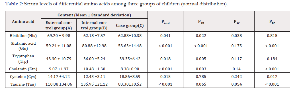
Table 2:Serum levels of differential amino acids among three groups of children (normal distribution).
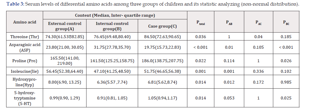
Table 3:Serum levels of differential amino acids among three groups of children and its statistic analyzing (non-normal distribution).
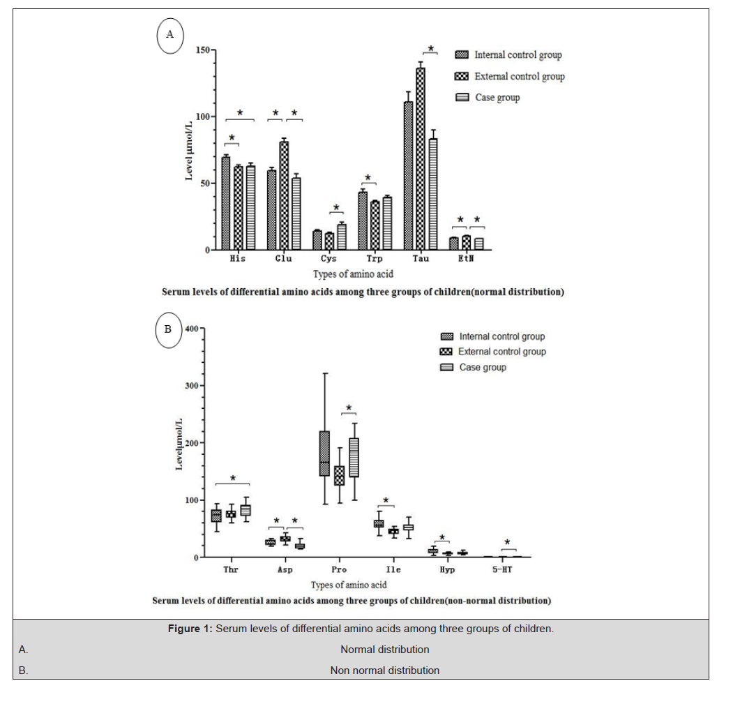
Figure 1:Serum levels of differential amino acids among three groups of children.
A. Normal distribution
B. Non normal distribution
Discussion
AAs metabolism is associated with many diseases [10]. Under normal physiological conditions, the concentration of AAs and their metabolites are maintained at stable levels [11]. Thus, it is critical to detect any change of these molecules to predict disease insurgence and course [12]. Change in AAs can also affect body growth and development. KBD is indeed linked several metabolic alterations in the body [13]. The detection of changes in AAs and/or their metabolites might be a useful biomarker for disease diagnosis such as KBD. Research has shown that the establishment and development of OA are associated with inflammation and alterations in AAs metabolism and profiles [14]. In this study, we determined the serum AAs of KBD adolescent employing UPLC/Q-TOF-MS technology, and found 12 different AAs and their metabolites, including His, Glu, Trp, Cys, Etn, Tau, Thr, ASP, Pro, Ile, Hyp, 5-HT. Among them, the concentration of 5 AAs and their metabolites (Glu, Etn, Cys, Tau and ASP) was either significantly higher or significantly lower in the serum of KBD children as compared with the controls (p<0.05). The serum concentrations of Hyp, Ile, and Trp between internal and external controls were significantly different (p<0.05). The concentrations of His, Thr, Proline, and 5-HT among the three groups were significantly different (p<0.05). These AAs and their metabolites were involved in several biological processes including aminoacyl-tRNA biosynthesis, histidine, arginine and proline metabolism, alanine, aspartate and glutamate metabolism, glycine, serine and threonine metabolism. ASP is a key driver of AA metabolism and energy metabolism. Glu is involved in histidine, arginine and proline metabolism, alanine, aspartate and glutamate metabolism. His, Cys, Thr, and Pro are also involved in AA metabolism. These processes link amino acid metabolism with carbohydrate metabolism through the exchange of sugar [6]. What’s more, some final metabolites have the potential function of the biomarkers of KBD.
While the etiology of KBD remains elusive, three major environmental factors have been identified as being associated with this disease: selenium deficiency, grain contamination by mycotoxin- producing fungi, and water contamination by organic materials such as fulvic acid [15]. The main pathophysiological changes of KBD include chondrocyte necrosis, ageing and dedifferentiation, type II collagen degradation and loss of proteoglycans [16]. Glu and Cys are involved in synthesis of GSH. GSH, belonging to selenoproteins, plays a very important role in the pathogenesis of KBD [17]. Decreased selenoproteins could result in apoptosis and necrosis of chondrocytes, which played a crucial role in the pathogenesis of KBD [18]. Mycotoxin contamination (e.g., T-2 toxin) of locally produced cereals may increase the levels of reactive oxygen species and free radicals in the body, which may damage chondrocytes, disturb the extracellular matrix and induce excessive apoptosis and necrosis of chondrocytes in KBD patients [18]. Etn indicated the abnormality of lipid metabolism and oxidative stress [19]. Trp plays an important role in regulating the autoimmune [20]. 5-HT, a neurotransmitter, was formed by Trp [21]. Ile might also be effective to attenuate the progression of OA by inhibiting NO synthesis [22]. His can be converted to intermediates of the Tricarboxylic Acid (TCA) cycle. His, as well as their relevant metabolic pathways, might undergo pathological alterations during the development of OA [23]. Thr is reported to regulate epithelial cell migration and proliferation, cell differentiation, restoration of epithelial barrier functions, modulation of cell apoptosis and proteoglycan synthesis [24]. Moreover, Tau, a sulfur-containing AA, exists in the body in free form and, while not directly involved in protein synthesis, it regulates the metabolism of AAs. Taurine plays an important role in bone metabolism, can promotes the production of osteoblasts [25] and inhibits the formation of osteoclasts [26]. Tau could promote chondrocyte proliferation, maintain its phenotype, and increase the expression of type II collagen [27]. Knee OA cartilage is characterized by a decrease in Asp-RR, possibly due to increase in collagen type II synthesis [28]. Pro are the main components of type II collagen in cartilage. The increase of Pro suggested that type II collagen could have been partly broken down [29], which might aggravate cartilage degeneration.
Because of the low number of KBD cases, one of the limitations of this study is the small sample size. Nevertheless, this investigation provided new clues into the KBD pathogenesis. Biological relevance of these AAs on KBD developing and occurrence were unclear. Many genes encoding enzymes involved AAs metabolism were up-regulated during the KBD development. Further research is needed to probe the AAs changes in KBD using large size and investigating the effect on the metabolic pathways linked to these AAs. In summary, among the 50 AAs detected using UPLC-QTOFMS, 12 different AAs and their metabolites were found difference in serum of KBD adolescent. These AAs changes provide a new clue for KBD pathogenesis.
Statement of Ethics
The experimental protocol was approved by the Ethics Committee of Harbin Medical University (HMUMER2013-26). Study population was in accordance with the ethical principles outlined in the world medical association Helsinki’s declaration. An otal of 122 adolescents were recruited in this study, and their parents or guardians all gave their written informed consent.
Funding Sources
This work was supported financially by the national natural science foundation of China (No.81372937), basic research projects of Qinghai province(2017-ZJ-770) and the training program of natural science and engineering technology discipline leader of Qinghai province “Kunlun Talents, Science and Technology leaders” in 2023.
Conflict of Interest
There are no conflicts of interest.
Author Contributions
Qiang Li and Qiong Wang wrote this paper and carried out epidemical
investigation
and the statistical analysis.
Xin Zhou, Hongmei Xue, Jianling Wang, Jiquan Li were carried
out the sample’s selection
Lihua Wang and Zhijun Zhao conceived the whole study and
participated in its design and coordination, and helped to draft the
manuscript.
All authors read and approved the final manuscript.
Data Availability Statement
All data generated or analyzed during this study are included in this article [and/or] its supplementary material files. Further enquiries can be directed to the corresponding author.
References
- Duan C, Guo X, Zhang XD, Han Jie Yu, Hua Yan, et al. (2010) Comparative analysis of gene expression profiles between primary knee osteoarthritis and an osteoarthritis endemic to Northwestern China, Kashin-Beck disease. Arthritis Rheum 62(3): 771-780.
- Zhang Y, He Y, Zhang D, Meng Zhang, Mengying Wang, et al. Death of chondrocytes in Kashin-Beck disease: Apoptosis, necrosis or necroptosis? Int J Exp Path 99(6): 312‐322.
- Zignego DL, Hilmer JK, June RK (2015) Mechanotransduction in primary human osteoarthritic chondrocytes is mediated by metabolism of energy, lipids, and amino acids. J Biomech 48(16): 4253-4261.
- Zhao ZJ, Li Q, Yang PZ, Wang H, Kong LC, et al. (2013) Selenium: a protective factor for Kashin-Beck disease in Qing-Tibet plateau. Biol Trace Elem Res 153: 1-4.
- Haubruge E, Chasseur C, Debouck C, Begaux F, Suetens C, et al. (2001) The prevalence of mycotoxins in Kashin-Beck disease. Int Orthop (SICOT) 25(3): 159-161.
- Peng A, Wang WH, Wang CX, Wang ZJ, Rui HF, et al. (1999) The role of humic substances in drinking water in Kashin-Beck disease in china. Environ Health Perspect 107(4): 293-296.
- Adams SB, Setton LA, Nettles DL (2013) The role of metabolomics in osteoarthritis research. J Am Acad Orthop Surg 21(1): 63-64.
- Bouatra S, Aziat F, Mandal R, Guo AC, Wilson MR, et al. (2013) The human urine metabolome. PLOS ONE 8(9): e73076.
- Tang X, Wang S, Zhan S, Niu J, Tao K, et al. (2016) The prevalence of symptomatic knee osteoarthritis in china: results from the china health and retirement longitudinal study. Arthritis Rheumatol 68(3): 648-653.
- Zhu L, Zhao ZJ, Liu SY, Wang WY, Hu J, et al. (2016) Urinary metabolic profiling of rats with articular cartilage damage induced by T-2 toxin. Chin J Endemiol 35: 350-353.
- Wang YM, Liu JW, Zhang RY, Wang WY, Hu J, (2018) Quantitative analysis of serum amino acid spectrum in osteoarthritis rat. Chin J Endemiol 37(11): 863-868.
- Priori R, Scrivo R, Brandt J, Valerio M, Casadei L, (2013) Metabolomics in rheumatic diseases: the potential of an emerging methodology for improved patient diagnosis, prognosis, and treatment efficacy. Autoimmunity Reviews12(10): 1022-1030.
- Wu CY, Guo X (2012) The research progression of metabonomics on Kashin-Beck disease. Foreign Medical Science Section of Medgeography 33: 76-786.
- Wang Y M, Wang W Y, Sun L Y, Qiang Li, Zhi Jun Zhao, et al. (2020) Serum metabolomic indicates potential biomarkers and metabolic pathways of pediatric Kashin-Beck disease [J]. Biomed Environ Sci 33(10): 750-759.
- Guo X, Ma WJ, Zhang F, Ren FL, Qu CJ, et al. (2014) Recent advances in the research of an endemic osteochondropathy in China: Kashin-Beck disease, Osteoarthritis and Cartilage 22(11): 1774-1783
- Zhang QM, Li H, Zhang ZD, Yang F, Chen JY (2015) Serum metabolites as potential biomarkers for diagnosis of knee osteoarthritis. Disease Markers e 684794.
- Wang SJ, Guo X, Zhang ZT, Guo R, Chen JH, et al. (2006) The apoptotic chondrocytes in articular cartilage of children and adults with Kashin-Beck disease. J Xi'an Jiaotong Univ Med Sci 27(6): 561e5.
- Chen JH, Xue S, Li S, Wang ZL, Yang H, et al. (2012) damage in Kashin-Beck disease and a rat Kashin‑Beck disease model by employing T-2 toxin treatment under selenium deficient conditions. J Orthop Res 30(8): 1229-1237.
- Zhao T, Yin TT, Zhang YZ, Lu J, Li P, et al. (2013) Study on common features of Damp-heat syndrome in rheumatism based on metabolomics. J Traditional Chinese Medicine 54(7): 592-596.
- Chase HW, Crockett MJ, Msetfi RM, Murphy R A, Clark L, et al. (2011) 5-HT modulation by acute tryptophan depletion of human instrumental contingency judgements. Psychopharmacology 213: 615-623.
- Sleight AJ, Marsden CA, Martin KF, Palfreyman MG (1988) Relationship between extracellular 5-hydroxytryptamine and behaviour following monoamine oxidase inhibition and L-tryptophan. Br J. Pharmacol 93(2): 303-310.
- Yang Y, Wu ZL, Meininger CJ, Guoyao Wu (2015) l-Leucine and NO-mediated cardiovascular function. Amino Acids 47: 435-447
- Li YS, Xiao WF, Luo W, Zeng C, Deng ZH, et al. (2016) Alterations of amino acid metabolism in osteoarthritis: its implications for nutrition and health. Amino Acids 48(4): 907-914.
- Lan A, Blachier F, Benamouzig R, Martin Beaumont, Christophe Barrat, et al. (2015) Mucosal healing in inflammatory bowel diseases: is there a place for nutritional supplementation? Inflamm Bowel Dis 21(1): 198-207.
- Park S, Kim H, Kim SJ (2001) Stimulation of ERK2 by taurine with enhanced alkaline phosphatase activity and collagen synthesis in osteoblast-like UMR-106 cells. Biochem Pharmacol 62(8): 1107-1111.
- Koide M, Okahashi N, Tanaka R, K Kazuno, K Shibasaki, et al. (1999) Inhibition of experimental bone resorption and osteoclast formation and survival by 2-aminoethanesulphonic acid. Arch Oral Biol 44(9): 711-719.
- Liu Q, Lu Z, Wu H, Zheng L (2015) Chondroprotective effects of taurine in primary cultures of human articular chondrocytes. Tohoku J Exp Med 235(3): 201-213.
- Catterall JB, Zura RD, Bolognesi MP, Kraus VB (2016) Aspartic acid racemization reveals a high turnover state in knee compared with hip osteoarthritic cartilage. Osteoarthritis Cartilage 24(2): 374-381.
- Xu ZW, Chen TM, Luo J, Ding SJ, Gao SC, et al. (2017) Cartilaginous Metabolomic study reveals potential mechanisms of osteophyte formation in osteoarthritis. J Proteome Res 16(4): 1425-1435.




 We use cookies to ensure you get the best experience on our website.