
Research Article 
 Creative Commons, CC-BY
Resistance of the Cranial and Caudal Cruciate Ligaments in Dogs (Canis lupus familiaris Linnaeus, 1758)
*Corresponding author:Thalyta Thayanny Santos Souza, Veterinary Medicine student, Federal University of Vale do São Francisco (Univasf), Avenida José de Sá Maniçoba, s/n°, Centro, Petrolina, Pernambuco, CEP-56304-917, Brazil.
Received:September 26, 2025; Published:October 01, 2025
DOI: 10.34297/AJBSR.2025.28.003716
Abstract
This study aimed to biomechanically characterize the Cranial Cruciate Ligament (CrCL) and Caudal Cruciate Ligament (CaCL) of dogs through destructive tensile mechanical tests, correlating the results with body biometric variables. A total of 40 ligaments from 10 adult, mixed-breed dogs were used, subjected to mechanical testing in a universal testing machine. The parameters of maximum strength (N), maximum deformation (mm), and stiffness (N/mm) were evaluated, as well as correlations between antimers and between the two types of ligaments. The results showed that the CaCLs exhibited greater strength and deformation compared to the CrCLs, with males standing out by showing higher values in almost all parameters. No statistically significant correlation was found between body measurements and the biomechanical parameters of the ligaments. Bone fracture was observed in 25% of the samples before ligament rupture, indicating the influence of the fixation technique and testing speed. The internal correlations between the ligaments suggest a synergistic role in joint stability. It is concluded that the caudal cruciate ligaments have greater biomechanical capacity than the cranial ones, and that intrinsic structural factors are more determinant than biometric variables. The data obtained provide support for the development of ligament substitutes and the improvement of surgical techniques in veterinary medicine.
Keywords:Ligament biomechanics; Mechanical traction; Body biometrics; Knee.
Introduction
The rupture of the Cranial Cruciate Ligament (CrCL) is the most common ligament injury in the stifle joint of dogs, being responsible for instability and progressive joint degeneration. The CrCL prevents cranial displacement of the tibia relative to the femur, in addition to controlling internal rotation and hyperextension of the joint [27]. Rupture of the Caudal Cruciate Ligament (CaCL) is less frequent and generally associated with concomitant injury to the CrCL [27,12].
Recent biomechanical studies have deepened the understanding of the femorotibial joint by using computational models based on finite elements to simulate the forces involved in injury and stabilization of the stifle [16]. Contemporary reviews highlight the multifactorial etiology of CrCL rupture, involving biomechanical, genetic, and environmental factors, in addition to emphasizing the integration of surgical techniques and rehabilitation protocols to optimize functional recovery [30].
Several surgical techniques are available for joint stabilization after CrCL rupture, including intracapsular and extracapsular methods, fibular head transposition, and tibial plateau leveling osteotomies [17]. Advances in biomechanical testing have enabled the quantification of the mechanical properties of ligaments, such as strength and elasticity, which are essential for evaluating treatment effectiveness [9,27]. Understanding the biological and biomechanical aspects of ligament pathologies is vital for improving diagnoses, preventive strategies, and therapies in dogs [6].
The canine stifle joint is a complex structure composed primarily of the femur, tibia, and fibula, as well as intra-articular menisci that correct incongruities between the articular surfaces [9,18]. This synovial joint is enclosed by a capsule that produces synovial fluid, essential for the lubrication and nutrition of the articular cartilage [29].
The main ligaments include the medial and lateral collateral ligaments, and the cranial and caudal cruciate ligaments, which ensure stability and allow flexion, extension, and limited rotational and translational movements [22,5]. The cruciate ligaments interlace, providing rotational stability and limiting hyperextension [1]. The Cranial Cruciate Ligament (CrCL) has a craniomedial portion that remains tense throughout the entire range of motion, and a caudolateral portion whose tension varies according to the degree of stifle flexion [13]. Microscopically, ligaments are composed of collagen bundles grouped into fascicles, primarily supplied by synovial tissues, with a central zone that is less vascularized, which may influence their susceptibility to injury [28]. Ligament innervation enables proprioceptive reflexes that help prevent excessive movements [29].
Cranial Cruciate Ligament (CCL) rupture typically occurs due to internal rotation forces and hyperextension of the stifle joint, especially during abrupt movements and fixed weight-bearing of the limb [12]. Factors such as aging, obesity, and genetic predisposition influence the progression of the injury, which leads to joint instability, osteoarthritis, and meniscal involvement [31,2]. Large breeds, such as Rottweilers and Labrador Retrievers, are more predisposed, especially in young individuals [21].
The diagnosis is confirmed by the cranial drawer test, which identifies abnormal tibial displacement, although partial and chronic lesions may complicate the assessment [29,12]. Caudal Cruciate Ligament (LCCa) injuries are rare and often traumatic, with a lower functional impact [15].
Uniaxial tensile tests are widely used to evaluate ligament strength, characterizing the relationship between applied load and deformation until rupture [9]. Ligaments exhibit viscoelastic, anisotropic, and nonlinear behavior, adapting to different directions and types of load [33]. During tension, collagen fibers initially stretch and align, followed by a linear phase until progressive rupture of the fascicles and tissue collapse, at which point the maximum load is reached [4]. Recent studies indicate that histological changes precede ligament failure, including extracellular matrix remodeling and cellular apoptosis, emphasizing the importance of early diagnosis [23].
In light of the above, the present study aimed to determine the maximum strength and deformation capacity of the cranial and caudal cruciate ligaments in dogs.
Materials and Methods
Material Acquisition
With the approval of the Human and Animal Studies Ethics Committee under protocol No. 27051115, fifty-three adult dogs-26 males and 27 females, of no defined breed and medium size-were collected from the Zoonoses Control Center (CCZ) of the Municipality of Petrolina (PE). The animals, already euthanized, were sent to the Laboratory of Anatomy of Domestic and Wild Animals at the Federal University of the São Francisco Valley (Univasf).
Body Biometry
For the assessment of body biometry, a measuring tape and a caliper with millimeter precision were used to obtain the following measurements:
a) Height: measured from the distal end of the thoracic limb to
the shoulder region, corresponding to the spinous processes
of the thoracic vertebrae located between the scapulae, also
referred to as the withers.
b) Length: measured along the vertebral column from the nuchal
crest to the base of the tail.
c) Shoulder girdle width: measured between the contralateral
scapular spines.
d) Pelvic girdle width: measured between the coxal tuberosities
of the iliac wings.
e) Thoracic height: measured from the base of the sternum to
the spinous processes of the seventh or eighth thoracic vertebrae.
f) Abdominal height: measured from the umbilical scar to the
spinous processes of the third or fourth lumbar vertebrae.
g) Cranial length: measured from the frontonasal suture to the
nuchal crest.
h) Nasal length: measured from the tip of the nose to the frontonasal
suture.
i) Head width: measured between the lateral surfaces of the
contralateral zygomatic arches.
j) Head height: measured from the nuchal crest to the mandibular
angle.
Body mass was determined using a digital scale, with values expressed in kilograms.
Dissection of the Material
After completing the body biometry of the cadavers, the pelvic limbs were first disarticulated at the coxal joint using a scalpel and forceps. The limbs were dissected by reflecting the muscles of the femoral region, as well as other adjacent structures such as ligaments, vessels, and nerves. The same procedure was performed in the tibial region: muscles covering the tibia were reflected, vasculonervous structures were sectioned, and adipose and connective tissues were removed to expose the tibia, leaving only the femur and tibia with the joint intact. Subsequently, in the femorotibial joint, the joint capsule, synovial bursae, and ligaments not relevant to the experiment were reflected.
After completing the dissection, only the femorotibial joint with intact cranial and caudal cruciate ligaments was obtained. After finishing the dissection, the femur and tibia were fractured at their midshaft using a band saw (Figure 1).
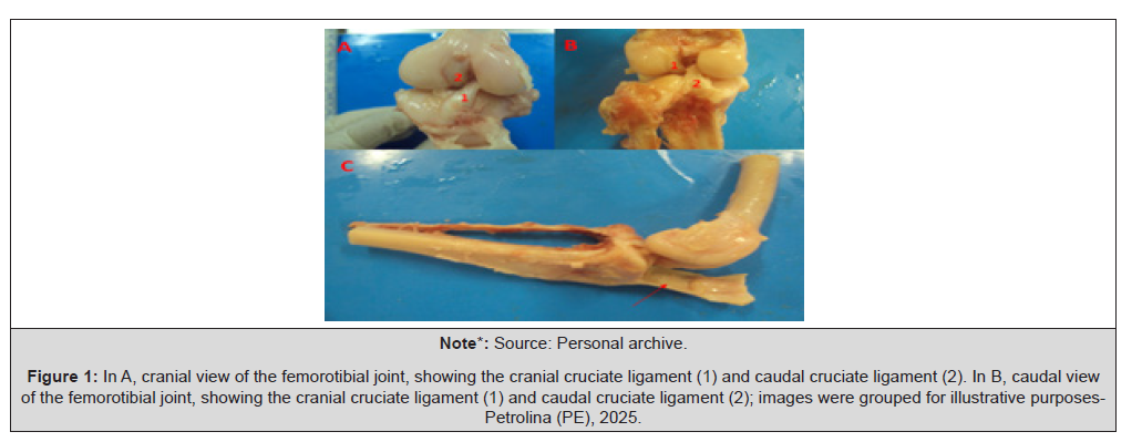
Figure 1:In A, cranial view of the femorotibial joint, showing the cranial cruciate ligament (1) and caudal cruciate ligament (2). In B, caudal view of the femorotibial joint, showing the cranial cruciate ligament (1) and caudal cruciate ligament (2); images were grouped for illustrative purposes- Petrolina (PE), 2025.
It is important to emphasize that during the dissection of the joints, if any articular or bone abnormalities were detected, the sample was discarded, as such conditions could interfere with the experimental results.
Ligament Biometry
After dissection of the joints, the medio-lateral and cranio-caudal thicknesses of all ligaments involved in the study were measured using a caliper, in both antimeres.
Destructive Mechanical Testing
The joint specimens were taken to the Materials Testing Laboratory at Univasf, where the mechanical tests consisted of tensile testing of the cranial and caudal cruciate ligaments. For this purpose, the femur was secured to the upper grip and the tibia to the lower grip (Figure 2). Once fixed, the machine was operated via computer, initiating traction on the joint until the ligament no longer resisted the applied force, thereby recording the maximum load (N) and deformation (mm) (Figure 2).
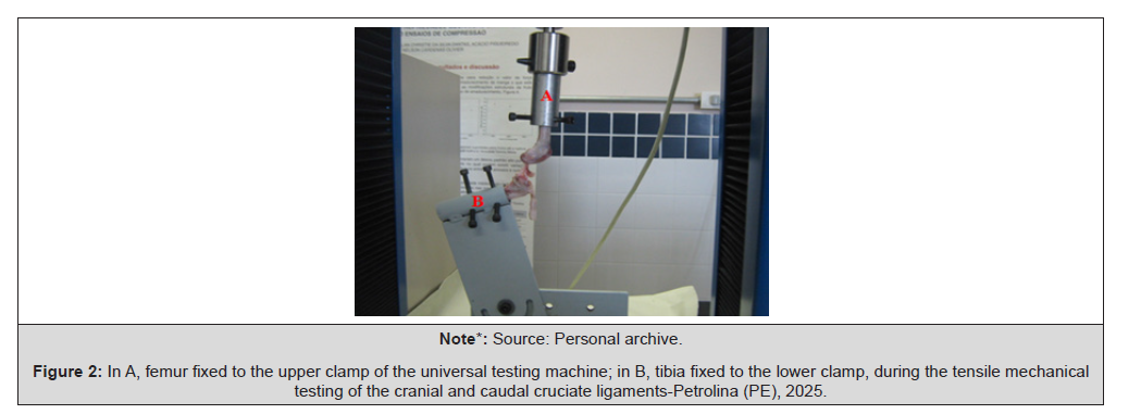
Figure 2:In A, femur fixed to the upper clamp of the universal testing machine; in B, tibia fixed to the lower clamp, during the tensile mechanical testing of the cranial and caudal cruciate ligaments-Petrolina (PE), 2025.
Statistical Analysis
Initially, the data were tabulated in Excel, dividing the samples into two groups according to sex. Each variable underwent a preliminary analysis using the R program to detect possible outliers. After this preliminary work, statistical analysis was performed using Student’s t-test in SAS 9.2, adopting correlations with a significance level greater than 0.7.
Results and Discussion
The means, standard deviations, and confidence intervals of the body biometric data of females and males are described in Table 1. The body biometric data showed significant differences between sexes, with males presenting higher values in all evaluated parameters. The 95% confidence intervals indicated consistency in the morphological differences between sexes, suggesting that these variations may directly influence biomechanical and functional aspects, such as the distribution of joint forces and the resistance of ligamentous tissues.
Consulting the literature regarding the biomechanical analysis of the ligaments studied in this work, it was found that these do not report the body biometric data of the animals used; or, when mentioned, it is limited to body mass, as reported by Brendolan, et al; (2001) [3], who studied the mechanical properties of the fascia lata and the cranial cruciate ligament, and reported only the body mass of the dogs, which averaged 11.8 kg±1.99-values close to those observed in this study. [20] Las Casas, et. al; (2008), when analyzing the mechanical properties of the cruciate ligaments, reported that the dogs used weighed between 34kg and 36kg, representing a range higher than that of the animals in this experiment. The performance of body biometry in the dogs used in the experiment is important for establishing possible relationships between ligament resistance and body dimensions-such as height, length, and circumferences- which varied significantly between males and females, highlighting the relevance of morphological characterization for biomechanical studies (Table 1).
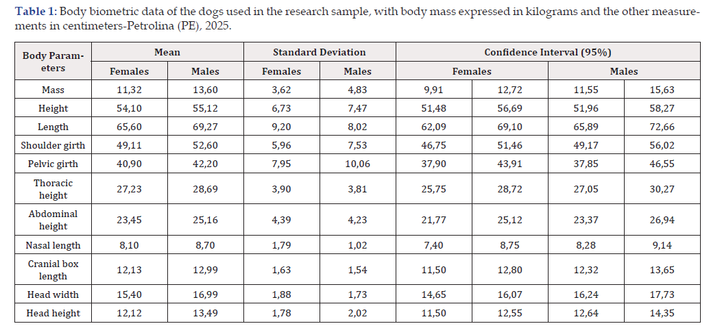
Table 1:Body biometric data of the dogs used in the research sample, with body mass expressed in kilograms and the other measurements in centimeters-Petrolina (PE), 2025.
Note*: Source: Author’s own work.
In the present study, the cranio-caudal and medio-lateral thicknesses of the ligaments were measured at their mid-third. Table 2 presents the biometric data of the cranial and caudal cruciate ligaments of the dogs used in the research, with measurements expressed in millimeters. It was observed that males exhibited higher mean values in all ligament dimensions compared to females, for both the CrCL and CaCL. In the CrCL, the left medio-lateral thickness was the largest among the evaluated parameters, averaging 3.87mm in males and 3.21mm in females. The cranio-caudal thickness was also greater in males, especially on the left side (2.66mm) compared to females (2.17mm). In the CaCL, males presented a left medio-lateral thickness of 3.21mm, while females recorded 2.75mm. The smallest measurement was observed in the right cranio- caudal thickness of the caudal ligament in females (2.03mm). Standard deviations indicate greater variability in male measurements, particularly in the right CrCL medio-lateral thickness (SD=1.19 mm). These findings suggest that ligament dimensions vary according to sex, limb side, and ligament type, which may directly influence their biomechanical resistance and structural behavior under joint loads (Table 2).
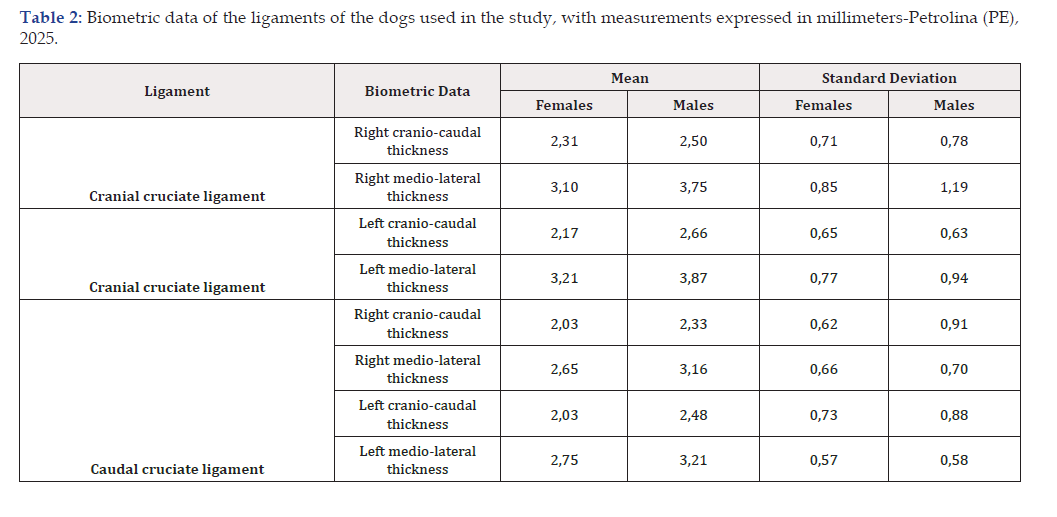
Table 2:Biometric data of the ligaments of the dogs used in the study, with measurements expressed in millimeters-Petrolina (PE), 2025.
Note*: Source: Author’s own work.
Fifty-three dogs were used in the experiment, totaling 106 joints subjected to destructive tensile mechanical testing. Among these, 29 joints (29.35%) presented bone fractures prior to ligament rupture, indicating structural failure at the bony ends during the test. The fractures occurred predominantly in the femoral condyles, the proximal tibial epiphysis, or at the insertion sites of the cruciate ligaments (Figure 3).
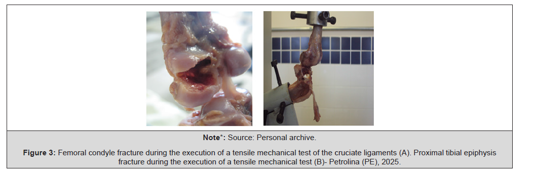
Figure 3:Femoral condyle fracture during the execution of a tensile mechanical test of the cruciate ligaments (A). Proximal tibial epiphysis fracture during the execution of a tensile mechanical test (B)- Petrolina (PE), 2025.
The probable cause of these fractures was attributed to the initial method of securing the bones in the grips of the testing machine, as the ruptures occurred when the bone segments were fixed at their extremities. After adjusting the technique, with fixation closer to the joint, the problem was resolved. [20] Las Casas, et .al; (2008), when studying the mechanical properties of the cruciate ligaments, reported a similar occurrence of femoral epiphyseal fractures during the first attempts, which was resolved after adjustment of the fixation.
[7] Costa, et. al; (2007) also observed bone fractures prior to Cranial Cruciate Ligament (CCL) rupture in pigs, reinforcing the importance of fixation technique in tensile tests. [3] Brendolan, et. al; (2001) reported only one avulsion of the lateral femoral condyle before ligament rupture, associating the event with the animal’s bone immaturity, as dentition indicated an approximate age of one year, possibly with growth plates still open. The influence of age on ligament resistance remains controversial. Haut, Lancaster, and [12] Decamp, et. al; (1992) concluded that the failure load of the patellar ligament does not vary with age. Conversely, [31] Vasseur, et. al; (2007) states that the resistance of the cranial cruciate ligament in dogs decreases with aging, attributing this reduction to progressive disorganization of collagen bundles (Figure 3).
Another relevant factor that can influence ligament strength and predispose to bone fracture prior to rupture is the sensitivity of the structural properties of the bone-ligament-bone complex to the rate of deformation applied during testing. Studies have shown that at high speeds, ligaments tend to fail under higher loads, with failure occurring predominantly in the ligament portion. Conversely, at lower speeds, there is a greater propensity for bone avulsion [24,8]. This behavior is characteristic of viscoelastic tissues, such as ligaments and tendons, which respond differently depending on the deformation rate.
During the tests conducted, it was observed that the Cranial Cruciate Ligament (CCL) ruptured before the Caudal Cruciate Ligament (CaCL), suggesting greater biomechanical resistance of the CaCL, which may explain the higher clinical prevalence of CCL ruptures [25]. This difference is related to the intrinsic properties of the ligament complex and is relevant for diagnosis and the definition of therapeutic strategies in cases of joint instability.
Both ligaments exhibited a preferential rupture pattern in the middle third (Figure 4), a region subjected to higher mechanical stress due to the crossing of functional fiber bundles and lower local vascularization, contributing to their structural vulnerability [3,32]. Additionally, cellular and structural alterations, such as disorganization of collagen fibers, tenocyte apoptosis, and modulation of hormonal receptors-including those for relaxin-interfere with the homeostasis of the extracellular matrix, constituting early markers of ligament degeneration [26,23] (Figure 4).
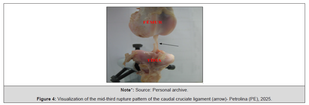
Figure 4:Visualization of the mid-third rupture pattern of the caudal cruciate ligament (arrow)- Petrolina (PE), 2025.
[20] Las Casas, et. al; (2008) emphasize that several factors can influence the results obtained in uniaxial tensile tests, as the methodology described in the literature shows considerable variation, especially regarding the method of structure fixation. Beyond technical differences, there is also inherent variability in biological tissues, whose mechanical properties may differ between individuals even under similar experimental conditions. This combination of factors contributes to discrepancies in results between studies, making direct comparisons and standardization of biomechanical parameters challenging. The mean values of maximum strength and deformation of the cranial and caudal cruciate ligaments, obtained through biomechanical tests, are presented in Table 3. It was observed that the caudal cruciate ligaments, both right and left, exhibited greater strength capacity compared to the cranial cruciate ligaments in both sexes. In the male group, the right CCLa showed the highest mean strength value (1882.90 ± 1670.60 N), followed by the right CCLr (1678.70±1628.60 N). Among females, the highest value was observed in the left CCLa (1384.50±733.60 N) (Table 3).
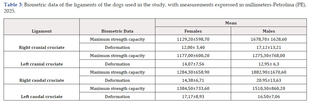
Table 3:Biometric data of the ligaments of the dogs used in the study, with measurements expressed in millimeters-Petrolina (PE), 2025.
Note*: Source: Author’s own work.
The comparative analysis with previous studies shows that the standard deviations observed in this work, although high, are consistent with those reported in the literature, even when methodological differences exist. Brendolan, Rezende and Pereira (2001), when performing tensile tests on the cranial cruciate ligament (CCL), obtained an average strength of 679.43 N±252.47 and an average deformation of 10.42 mm±2.61, values that correspond to approximately half the strength observed in this study, although the deformation values are similar, albeit slightly lower. This discrepancy can be attributed to the testing speed, which in the cited study was 8.77mm/min, whereas in this work it was 25mm/min. [20] Las Casas, et. al; (2008) caution that comparisons between studies should be made carefully, as there is considerable variation in the loading rate, as well as differences in fixation methods and experimental conditions. Even among samples of the same species subjected to the same loading rate, biomechanical behavior can vary significantly. Recent studies reinforce that ligament response to cyclic testing depends strongly on the fixation system, load rate, and number of cycles, directly influencing failure patterns [10].
The variability of the obtained data may also be related to histological differences between the ligaments, such as the composition and orientation of collagen fibers, degree of vascularization, and presence of specific cellular markers [11]. Despite the extensive morphometric characterization performed in this study, no statistically significant correlation was observed between body biometric parameters and the biomechanical properties of the ligaments, in either males or females. These findings support finite element– based computational models, which indicate that the structural response of the joint to loads or implants is not directly related to body size, but rather to the tissue properties of the ligament [19], emphasizing the importance of individualized approaches in the treatment of ligament ruptures. Additionally, no biomechanical studies specifically addressing the caudal cruciate ligaments in dogs were found in the consulted literature, highlighting the originality and relevance of the data presented in this work.
Based on the means and respective standard deviations of the analyzed variables, possible relationships between biomechanical and biometric parameters were assessed. It was observed that, in both males and females, there was no statistically significant correlation between body biometric parameters and the maximum strength or deformation of the Cranial Cruciate Ligament (CCL) and caudal cruciate ligament (CaCL). However, in the female group, strong correlations (r>0.7) were identified between the strength of the right CCL and the following variables:
a) its corresponding deformation (r=0.75345).
b) strength of the left CCL (r=0.72997).
c) strength of the right CaCL (r=0.96922).
d) strength of the left CaCL (r=0.70539).
Still among the females, the strength of the left CCL showed a high correlation with its deformation (r=0.71333) and with the strength of the ipsilateral CaCL (r=0.96391). In the male group, the strength of the right CCL was strongly correlated with its deformation (r=0.89911) and with the strength of the left CaCL (r=0.98570). The left CCL, in turn, demonstrated a high correlation with its deformation (r=0.85273) and with the strength of the CaCL of the same limb (r=0.97172).
These correlations suggest a synergistic function between the cranial and caudal cruciate ligaments in maintaining knee joint stability. Ex vivo biomechanical evaluations indicate that the integrity of the CCL is essential for the collective performance of the ligament complex, and its rupture requires therapeutic interventions that restore quasi-isometric points [14], promoting biomechanical balance and improved clinical outcomes.
Conclusion
The present study significantly contributes to the understanding of the biomechanical properties of the cranial and caudal cruciate ligaments in dogs through destructive tensile mechanical testing. The results demonstrated that the caudal cruciate ligaments exhibited greater resistance and deformation capacity compared to the cranial cruciate ligaments, with male animals standing out, showing higher mean values in almost all evaluated parameters. Despite extensive body biometrics characterization, no statistically significant correlation was observed between body dimensions and ligament resistance or deformation, reinforcing that structural and histological factors are determinant in the biomechanical response.
Internal correlations between the ligaments, particularly between contralateral pairs and between resistance and deformation, highlight the synergistic function of the ligament complex in knee joint stability. The occurrence of bone fractures prior to ligament rupture in some samples underscores the importance of fixation technique and testing speed in conducting biomechanical assays. Furthermore, the lack of previous studies on the caudal cruciate ligaments in dogs reinforces the originality and relevance of the data presented.
These findings provide technical-scientific support for future research aimed at the development of biocompatible materials for ligament replacement, as well as for the improvement of surgical techniques and individualized therapeutic strategies in cases of ligament rupture or degeneration.
Acknowledgements
The authors thank the National Council for Scientific and Technological Development (CNPq) for providing the scientific initiation scholarship.
Conflict of Interest
The authors declare no conflict of interest.
References
- Amis AA (2012) The Functions of the fibre bundles of the anterior cruciate ligament in anterior drawer, rotational laxity and pivot shift. Knee Surg Sports Traumatol Arthrosc 20(4): 613-220.
- Anderson KL, Zulch H, O’neill DG, Meeson RL, Collins LM (2020) Risk factors for canine osteoarthritis and its predisposing arthropathies: a systematic review. Front Vet Sci 7: 220.
- Brendolan AP, Rezende CMF, Pereira MM (2001) Biomechanical properties of the fascia lata and cranial cruciate ligament of dogs. Braz Arch Vet Med Anim Sci 53(1): 231-240.
- Carlstedt CA (1987) Mechanical and chemical factors in tendon healing. Acta Orthop Scand 58(224): 7-75.
- Constantinescu GM (2005) Clinical anatomy of small animals. Rio de Janeiro. Guanab Koog 400.
- Cook JL (2010) Cranial cruciate ligament disease in dogs: biology versus biomechanics. Vet Sug 35(8): 270-277.
- Costa RB (2007) Mechanical properties of the anterior cruciate ligament in a porcine model. 2007. 39f. Undergraduate Thesis (Bachelor of Medicine)- Federal University of Santa Catarina, Florianó
- Dota AF, Zenaide MR, Demange MK, Camanho GL, Hernandez AJ (2007) Study of the mechanical properties of the posterior cruciate ligament and patellar tendon on fresh human cadavers after radiofrequency shrinkage. Acta Ortop Bras 15(3): 138-142.
- Garcia A, Spim JA, Santos C (2000) Materials testing. 2nd ed. Rio de Janeiro: LTC 247.
- Goin B, Buttin P, Lafon Y, Massenzio M, Viguier E, et al. (2022) Biomechanical cyclic loading test of a synthetic ligament fixation system used for intra-articular stabilization of deficient canine stifles. Open Vet J 12(3): 341-350.
- Gonera B, Wysiadecki G, Kurtys K, Brzezinski P, Borowski A, et al. (2023) Immunohistochemical analysis of the ligamentum mucosum is the key to understand its clinical usefulness. Ann Anat- Anatomischer Anzeiger 249: 152106.
- Haut RC, Lancaster RL, Decamp CE (1992) Mechanical properties of the canine patellar tendon: Some correlations with age and the content of collagen. J Biomech 25(2): 163-173.
- Heffron LE, Campbell JR (1978) Morphology, histology and functional anatomy of the canine cranial cruciate ligament. Vet Rec 102(13): 280-283.
- Hsu WR, Lin CC, Sun CY, Wu CH (2023) Ex vivo biomechanical evaluation of extracapsular stabilization with quasi-isometric points in canine cranial cruciate ligament-deficient stifles. BMC Vet Res 19(1): 93.
- Johnson AL, Olmstead ML (1987) Caudal cruciate ligament rupture: a retrospective analysis of 14 dogs. Vet Surg 16(3): 202-206.
- Kiapour A, Kiapour AM, Kaul V, Quatman CE, Wordeman SC, et al. (2013) Finite element model of the knee for investigation of injury mechanisms: development and validation. J Biomech Eng 136(1): 011002.
- Kim S E, et al. (2008) Tibial osteotomies for cranial cruciate ligament insufficiency in dogs. Vet Surg 37(2): 111-125.
- König HE, Liebich HG (2004) Anatomy of domestic animals: text and color atlas. Porto Alegre: Artmed 2.
- Lang ML, Lupke M, Gotz M, Volk HA, Klasen J, Harms O (2024) Biomechanical Evaluation of a Novel Ceramic Implant for Canine Cranial Cruciate Ligament Rupture Treatment: A Finite Element Analysis Approach. Animals 14(22): 3296-3296.
- Las Casas EB, Resende JL, Faria MTC, Saffar JME, Oliveira EA (2008) Determination of the mechanical properties of cruciate ligaments via uniaxial tensile tests. Braz J Biomechan 9(17): 73-80.
- Matera JM, Tatarunas AC, Oliveira RMD, Brugnaro M, Macchione RF (2007) Retrospective epidemiological study of dogs with cranial cruciate ligament rupture: 323 cases (1999 a 2005). Braz J Vet Res Animal Sci 44(4): 88-95.
- Miller ME, Evans HE, Delahunta A (2001) Guide to Dog Dissection, 5th ed. Rio de Janeiro: Guanabara Koogan: 250.
- Niebauer GW, Restucci B (2023) Etiopathogenesis of Canine Cruciate Ligament Disease: A Scoping Review. Animals 13(2): 187.
- Pioletti DP, Rokotomanana LR, Leyvras PF (1999) Strain rate effect on the mechanical behavior of the anterior cruciate ligament–bone complex. Med Eng Phy 21(2): 95-100.
- Readioff R, Geraghty B, Elsheikh A, Comerford E (2020) Viscoelastic characteristics of the canine cranial cruciate ligament complex at slow strain rates. Peer J 8: e10635.
- Restucci B, Sgadari M, Fatone G, Valle GD, Aragosa F, et al. (2022) Immunoexpression of Relaxin and Its Receptors in Stifle Joints of Dogs with Cranial Cruciate Ligament Disease. Animals 12(7): 819.
- Romano L, Martins PCA, Schmaedecke A, Saut JPE, Ferrigno CRA (2006) Biomechanical analysis of the intact knee and knee with rupture of the cranial cruciate ligament regarding the degree of cranial displacement and joint stiffness in dogs. Acta Cir Bras 21(1): 46-50.
- Rooster H, Bruin T, Van Bree H (2006) Morphologic and functional features of the canine cruciate ligaments. Vet Surg 35: 769-780.
- Schulz K (2008) Cranial cruciate ligament rupture. In: Fossum TW, Small Animal Surgery. 3rd ed., Rio de Janeiro: Elsevier: 1254-1275.
- Spinella G, Arcamone G, Valentini S (2021) Cranial cruciate ligament rupture in dogs: review on biomechanics, etiopathogenetic factors and rehabilitation. Vet Sci (8)9: 186.
- Vasseur DB (2007) Knee joint, In: Slatter D. Manual of small animal surgery. 3rd ed. São Paulo: Manole 2: 2090-2116.
- Vasseur PB, Arnoczky SP, Lau RE (1985) Correlative biomechanical and histologic study of the cranial cruciate ligament in dogs. Am J Vet Res 46(9): 1842-1854.
- Wilchez DFT (2012) Study of the mechanical behavior of ligaments and tendons using optical measurement. 2012. 108 f. Master’s Dissertation (Master in Mechanical Engineering)- Federal University of Santa Catarina, Florianó




 We use cookies to ensure you get the best experience on our website.