
Case Report 
 Creative Commons, CC-BY
Fibrotic and Emphysematous Backgrounds in Bilateral Covid-19 Pneumonia: a Suggestive Case Report at Computed Tomography
*Corresponding author:Francesco Messina, Unit of Radiology - Riuniti Hospital Azienda Ospedaliera Grande Ospedale Metropolitano “Bianchi– Melacrino–Morelli” Via Giuseppe Melacrino n.21, 89124 Reggio Calabria – Italy.
Received:September 22, 2025; Published:October 06, 2025
DOI: 10.34297/AJBSR.2025.28.003717
Abstract
Covid-19 pneumonia can rapidly lead to serious pulmonary complications associated with pneumonia, posing a delicate challenge for the clinicians, especially radiologists, who are committed to ensuring a timely and early diagnosis of the disease. Serial chest imaging enable clinicians to better monitor disease progression and identify potential complications early which may decrease the mortality and morbidity associated with Covid-19 pneumonia. In our case we describe a patient with bilateral Covid-19 pneumonia, that collaterally had bilateral emphysematous background and pulmonary fibrosis, documented at Computed Tomography (CT).
Keywords:Emphysematous Background Covid-19, Pneumonia, Computed Tomography
Background
Pneumonia is the most important manifestation of Covid-19 infection, whilst acute respiratory distress syndrome (ARDS) is the most severe sequela. Presence of pleural effusion, lung cavitation, fibrosis and emphysemas are associated with severe disease and often carry poorer prognosis [1].
Many studies and cases have reported common Covid-19 chest manifestation on chest x-ray (CXR) and CT [2-6]. The use of CT imaging in the diagnosis and follow-up had rapidly grown, and radiological patterns along the disease course are increasingly understood. Chest CT imaging has been demonstrated more sensitive than chest radiography to identify the manifestations of Covid-19 pneumonia, for the severity assessment and monitoring of the disease [7].
Case Presentation
We report a case of an 82-year-old man presented to the Emergency Department with fever, cough and dyspnea. Clinical exam ination revealed an elevated body temperature of 38°C, while laboratory blood tests revealed elevated neutrophil count, raised C-reactive protein; the naso- pharyngeal sampling was positive for SARS-CoV-2.
High Resolution CT scan of the chest was urgently done (Picture 1, a-c), in basal conditions, and the images so obtained were analyzed with a slice-thickness of 1.2 mm and MPR reconstructions (axial, sagittal, and oronal).
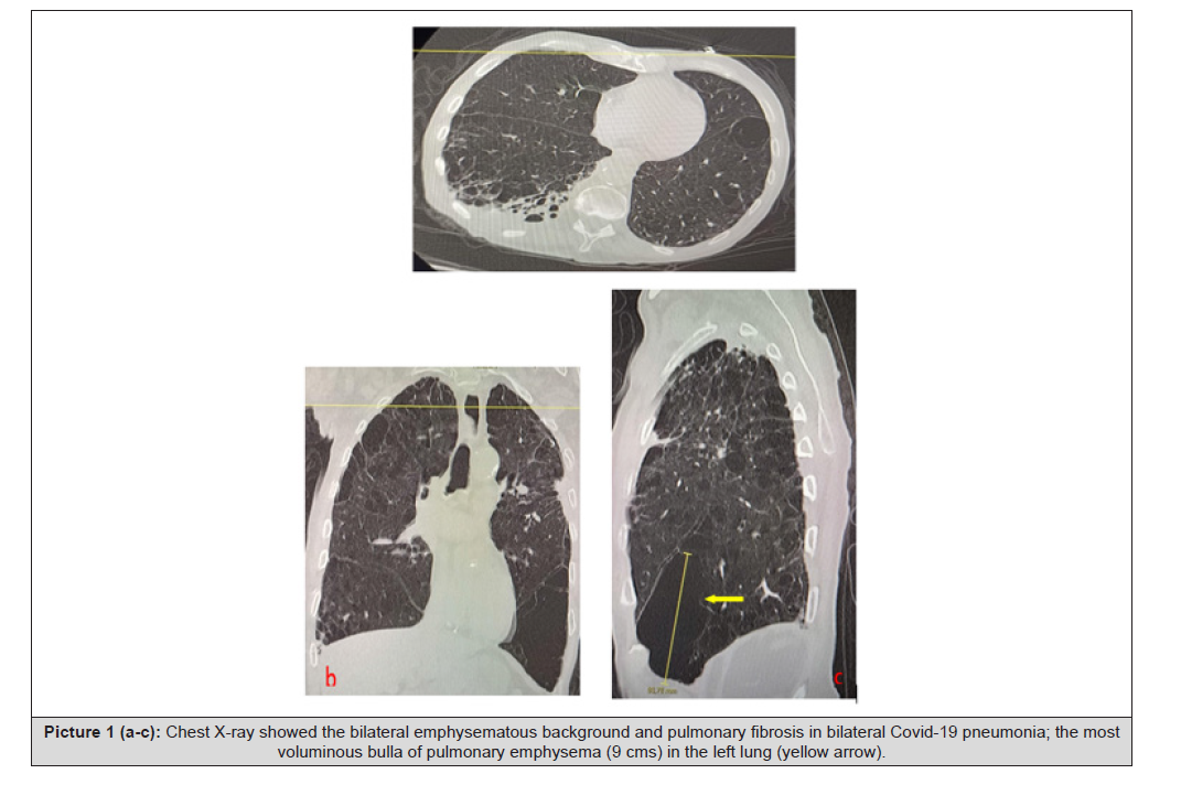
Figure 1 (a-c)::Chest X-ray showed the bilateral emphysematous background and pulmonary fibrosis in bilateral Covid-19 pneumonia; the most voluminous bulla of pulmonary emphysema (9 cms) in the left lung (yellow arrow).
CT had documented in both lungs the presence of a diffuse bilateral emphysematous background, characterised by the presence of multiple and diffuse bilateral pulmonary emphysema bullae, the most voluminous of them of about 9 cms, in the left lung. CT had also shown a bilateral fibrotic thickening of the pulmonary interstitium, also with thickened areas with a ground-glass pattern and interstitial thickenings. There were not pleural- pericardial effusions.
The patient was immediately hospitalized and treated and, after theree weeks of drug therapy, he had returned to home.
Discussion
Chest imaging is indicated in Covid-19 patients for establishing a baseline for the patient’s pulmonary condition, identification of other comorbidities and for monitoring the progression of disease. In the event of clinical deterioration, imaging assessment helps to diagnose disease progression and complications, such as pulmonary fibrosis, heart failure or vascular diseases. Typical chest findings in Covid-19 patients show bilateral lung involvement with patchy or asymmetric diffuse air space opacities, predominantly in a peripheral, posterior distribution and mainly in the lower lobes. In later stages of the disease, CT may show increased GGO, dispersed consolidation, reticular opacities, crazy-paving, bronchiectasis, pleural thickening, septal thickening and involvement of subpleural region. As the disease progresses, atypical CT features such as pleural and pericardial effusion, cystic changes, emphysematous riacutization, and nodules may be present [8-12].
Conclusion
Computed Tomography had an important role in the diagnosis and severity evaluation of Covid-19 pneumonia, because it investigates very well the dynamic CT changes in different stages of the disease, and also in the follow-up of the patients. CT scan can show a very important detection in the radiological alterations resembling a pattern of combined pulmonary Covid-19 pneumonia and pulmonary emphysema, with its background and other complications. Covid-19 pulmonary infection can leave a chronic impact on respiratory function, causing a clinical worsening of pre- existing conditions, such as emphysema and pulmonary fibrosis. On average, an adult may require from six to twelve months to recover, although this may not always be complete. This is caused by pulmonary fibrosis, which causes stiffening of the lung tissue affected by the infection, resulting in a further reduction in respiratory exchange functionality. A prompt recognition of these complications, a “target” ventilation and a drug therapy for the patients, and a follow-up strategy for post-Covid19 patients, are very important to evaluate the response to treatment and/or to document disease progression, making the direction of care for such patients and reduce their morbidity and mortality.
References
- Shi H, Han X, Jiang N, Cao Y, Alwalid O, et al (2020) Radiological f indings from 81 patients with COVID-19 pneumonia in Wuhan, China: A descriptive study. Lancet Infect Dis 4: 425-434.
- ACR recommendations for the use of chest radiography and computed tomography for suspected Covid-19 infection.
- Jin YH, Cai L, Cheng ZS, Cheng H, Deng T, et al (2020) A rapid advice guideline for the diagnosis and treatment of 2019 novel coronavirus (2019-nCoV) infected pneumonia (standard version). Mil Med Res 7(1): 4.
- Kanne JP, Little BP, Chung JH, Elicker BM and Ketai LH (2020) Essentials for radiologists on COVID- 19: an update. Radiology Scientific Expert Panel. Radiology 296(2): E113-E114.
- Messina F, Calabrese G, Turano L and Tebala C (2021) A pulmonary hamartoma in Covid-19 pneumonia: an interesting case studied with computed tomography. Radiol Case Rep 16(4): 942-944.
- Chan WY, Lim YT, Hamid MTR, Ng CC, Gowdh NFM, et al (2022) Pulmonary complications of Covid-19. Sultan Qaboos Univ Med J 22(1): 138-143.
- Messina F, Nucera A, Tripodi Ca and Arcadi N (2021) Pulmonary Emphysema in Covid-19 Pneumonia: CT Findings. International Journal of Clinical Studies and Medical Case Reports 10(4): 003.
- Nasir MU, Roberts J, Muller NL, Macri F, Mohammed MF et al. (2020) The role of emergency radiology in Covid-19: from preparedness to diagnosis. Can Assoc Radiol J 71(3): 293-300.
- Rubin GD, Ryerson CJ, Haramati LB, Sverzellati N, Kanne JP, et al (2020) The role of chest imaging in patient management during the Covid-19 pandemic: A multinational consensus statement from the Fleischner Society. Chest 158: 106-116.
- Sun R, Liu H and Wang X (2020) Mediastinal emphysema, giant bulla, and pneumothorax developed during the course of Covid-19 pneumonia. Korean J Radiol 21: 541-544.
- Salehi S, Abedi A, Balakrishnan S and Gholamrezanezhad A (2020) Corona- virus disease 2019 (Covid- 19): A systematic review of imaging findings in 919 patients. AJR Am J Roentgenol 215: 87-93.
- Messina F, Calabrese G, Tebala C and Turano L (2025) A Case of Pericardial Effusion in A Patient with Pace-Maker and Covid-19 Pneumonia at MSCT. Am J Biomed Sci & Res 28(2): 247-249.




 We use cookies to ensure you get the best experience on our website.