
Research Article 
 Creative Commons, CC-BY
Evaluation of Y Chromosome Azoospermia Factor (AZF) Microdeletion, Hormone Analysis and Testis Morphology in Different Infertile Males
*Corresponding author:Prof. A. S. Ansari, PhD, FAMS, Reproductive Health Laboratory, Centre for Advanced Studies, Department of Zoology, University of Rajasthan, Jaipur, India.
Received:August 18, 2025; Published:August 21, 2025
DOI: 10.34297/AJBSR.2025.28.003658
Abstract
Male infertility with the genetic factors might be the result of chromosomal abnormality and Y chromosome microdeletions. For this it is mandatory to evaluate carefully the karyotypes and AZF microdeletion prior to opt assisted reproductive techniques to reduce the genetic risk for the new generation. For the study 127 infertile subjects were recruited from the Division of Infertility, Department of Urology, SMS Medical College and Attached Hospitals, Jaipur, Rajasthan, India. Molecular analysis for AZF loci was carried out using multiplex (PCR). Each patient was examined for eight AZF loci that mapped to interval 5 and 6 of the Y chromosome. Semen characteristics and serum hormones were analyzed. Out of 127 subjects, 19.68% subjects were normozoospermic, 11.81% oligozoospermic, 21.25% severe oligozoospermic, 27.55% azoospermic and 19.68% were asthenozoospermic and 15 fertile (control) subjects were undertaken for this purpose. Yq Microdeletion was screened in 30 subjects out of 127 infertile subjects. The Yq microdeletion was present in 4 out of 30 infertile subjects (13.33%). Out of 8 azoospermic subjects one had microdeletion in AZF c region and one had microdeletion in AZF b region, 6.66% microdeletions were present in azoospermic group. Out of 6 asthenozoospermic subjects, one had microdeletion in AZF a region and one subject had microdeletion in AZF b region from teratozoospermic subject. No microdeletion in AZF was detected in other subjects. Serum hormone FSH level was nonsignificantly high, whereas, LH and prolactin levels were drastically (p<0.001) high in azoospermic subjects. Testosterone level was found nonsignificantly decreased in azoospermic subjects as compared to other subjects. No remarkable changes were present in PSA levels. Our findings support the implementation of genetic screening for Y chromosome microdeletions in infertile males undergoing assisted reproductive technologies (ART), to mitigate the risk of vertical transmission of Yq microdeletions to offspring conceived via ART or related methodologies.
Keywords:Male infertility, Yq Microdeletion, Serum Hormone, PSA, Testicular Morphology, Azoospemia
Abbreviations:AZF: Azoospermia Factor; PCR: Polymerase Chain Reaction; ICMART: International Committee That Monitors Assisted Reproductive Technologies; FSH: Follicle Stimulating Hormone; LH: Luteinizing Hormone; STS: Sequence Tagged Sites; IEC: Institute Ethical Committee; PBS: Phosphate Buffer Saline; SDS: Sodium Dodecyl Sulphate; FNAC: Fine Needle Aspiration Cytology; SD: Standard Deviation; SCOS: Sertoli Cell Only Syndrome; ART: Assisted Reproductive Technology; PSA: Prostate Specific Antigen
Introduction
In the mammalian system, the biological information is transferred from parents to offspring through the genetic material. Sperm DNA is known to contribute one half of the genomic material to offspring. Recently, Leslie, et al. [1] found that 20 % of infertility cases were attributed only to male factors. Thus, normal sperm genetic material is required for fertilization, embryo and fetal development and post-natal child well-being [2]. An international committee that monitors assisted reproductive technologies (ICMART) defines infertility is a medical disorder of incapability of a couple to conceive despite engaging in frequent, unprotected intercourse for over a period of twelve months [3]. Various in vivo and in vitro studies have suggested that disturbances in the organization of the genomic material in sperm nuclei are negatively correlated with the fertility potential of spermatozoa [4]. An appropriate endocrine milieu is also crucial for sexual differentiation, normal reproduction as well as maturation of spermatozoa. This is maintained through hypothalamo-hypophysis - gonadal axis and disturbance in this axis may result in infertility. Major hormones involved in this control are Follicle Stimulating Hormone (FSH), Testosterone and Luteinizing Hormone (LH). The LH secreted from the adenohypophysis is responsible for the proper androgenic environment. The other gonodotropic hormone, FSH affects germinal epithelium and Sertoli cells. Testosterone is responsible for the development of secondary characteristics. Therefore, FSH determination is very much important for examination of epithelial function of seminiferous tubules [5].
Biopsy is a standard procedure for the study of diseases of various organs. Testicular biopsy, after its first introduction [6] became a common procedure in the andrology laboratory to differentiate between spermatogenic failure and ductal obstruction. Since then, several patterns of testicular biopsy have been reported from time to time [7-12]. Testicular biopsy provides useful information in selection to diagnosis, prognosis, and choice of treatment in infertile men. After hormone analysis, testicular biopsy can be performed for further investigation. FSH is regarded as the main indicator for biopsy exploration. High FSH values discuss total germ cell failure or testicular damage and, therefore, biopsy is of no use in these cases [13-15]. A high FSH value is, thus compatible with primary testicular failure [16] and is usually found in subjects with Sertoli cell only syndrome and Klinefelter’s syndrome [17,18]. Encroachment research in molecular reproductive biology enabled the detection and characterization of fundamental genetic causes of male infertility. Affected infertile couples must be genetically sought by systematic evaluation of causes and informed about the implications of such diagnoses for assisted reproductive technology outcome and their impending progeny. An association between human male infertility and chromosomal anomalies has been known for a long time [19,20]. The potential association between chromosomal abnormalities and male infertility was first highlighted by the results of large-scale karyotype surveys conducted by Kjessler [21] and Chandley [19] in subfertile males.
The Y chromosome in mammals carries the gene that switches the development of the indifferent gonad from the default female pathway to the male pathway and results in the development of the testis [22]. Deletions in the long arm of Y chromosome (Yq) were suggested to be responsible cause for azoospermia [23]. The long arm of the Y chromosome contains genes and gene families involved in spermatogenesis and are important for germ cell development and differentiation. In 1996, Vogt and colleagues [24] have defined three non-overlapping regions for spermatogenesis loci to as azoospermia factor (AZF a, b and c from proximal to distal Yq).
Microdeletions in the AZF region of the Y chromosome represent one of the most prevalent genetic causes of male infertility, as spermatogenesis is regulated by numerous genes located on both the autosomes and the Y chromosome. Vollrath and colleagues [25] constructed a 43-interval deletion map of a human Y chromosome that contained an ordered array of sequence tagged sites (STS) which spanned the entire length of the Y chromosome. The genes vital for spermatogenesis are positioned on the long arm of the Y chromosome in deletion interval 5 and 6 bend 11.23. This region is referred to as the AZF as the most severe phenotype associated with its deletion is azoospermia.
The AZFa locus is located on proximal Yq11 (Yq 11.21), while AZFb and AZFc are located on distal Yq 11 (Yq 11.23). Deletions in the AZFc region might cause serious consequences such as spermatogenic arrest and is coupled with azoospermia Sertoli cell only syndrome to hypo-spermatogenesis to maturation arrest. Certain gene mutations linked with pathological syndromes also have a role in male infertility. Severe testicular disorders in infertile males often include deletions on the long arm of the Y chromosome [26,27]. The typical Y chromosome microdeletions for infertile males were 8.2% and majority of microdeletions (84.3%) were associated with azoospermia [28]. These deletions in fertile controls have been reported to be less than 1% and no deletion has been detected in men with normal semen analysis [29].
A significant proportion of couples in reproductive age suffer from primary and/or secondary infertility. Couples genetic counseling states about the genetic make-up and any involved risk factor that may be conferred to progeny. A variety of possible causes on male infertility, as mentioned above, have been identified in majority of cases, the diagnosis of male infertility revolves around the routine semen analysis. Therefore, incorporating cytogenetic analysis is crucial for diagnosing the specific type of male infertility and for preventing the transmission of genetic abnormalities to future generations, particularly in infertile couples pursuing ART following appropriate genetic counseling.
Materials and Methods
Subject Recruitment
Five hundred infertile couples were selected for initial screening. After exclusion of infertility through female factors, males above 45 years, aspermic patients, any known reproductive pathology (e.g. genital tract infections, prostatitis, epididymitis, etc.) or any hormonal therapy in the last six months and patients with erectile dysfunction, 127 subjects were primarily recruited for the present investigation from the Division of Infertility, Department of Urology, SMS Medical College and Attached Hospitals, Jaipur, Rajasthan, India. All subjects signed an informed consent form in order to undergo a full medical consultation, clinical examination, sample collection and relevant biochemical testing. Clinical examination of all subjects was carried out and information on age, health problems, history of infertility in the family, height, weight, etc. were recorded. The study was approved by the Institute Ethical Committee (IEC).
Collection of Semen Samples
Semen samples were collected by masturbation into a clean sterile sample collection container, under aseptic conditions. Subjects were instructed to abstain for at least 48 hours prior to collection the semen sample. The samples were liquefied for at least 20 minutes in a water bath at 37 °C, but no longer than 1 hour prior to performing a routine semen analysis.
Semen Characteristics
Semen analysis was performed within one hour after collection of semen. Analyses were performed to assess semen liquefaction, color, odor, pH, appearance, consistency, agglutination, semen volume, sperm motility, vitality, morphology, and sperm concentration [30].
Azoospermia Factor (AZF) Microdeletion Analysis
Out of 127 infertile subjects 30 infertile subjects were studied for AZF microdeletion and 15 fertile (control) subjects were used for the purpose. Molecular analysis for AZF loci was carried out using PCR. Each patient was examined for eight AZF loci that mapped to interval 5 and 6 of the Y chromosome. The SRY sex determining region on the short arm of Y chromosome (SY 14) was used as interval control.
Isolation of DNA from Sperm
The sperm DNA was isolated according to the method of Laird, et al. [31]. In brief, the sperm was separated from seminal plasma by centrifugation at 300 g for 10 min. and washed with phosphate buffer saline (PBS). The obtained sperm pellet was lysed after adjusting the sperm concentration to 30×10⁶ to 120×10⁶ sperm/5 mL in lysis buffer containing 100 mM Tris-HCl, 5 M EDTA, 0.2% sodium dodecyl sulfate (SDS), 200 mM NaCl, 100 μg proteinase K, and 80 mM dithiothreitol (DTT). The mixture was freshly prepared and incubated at 37 °C for 24 hours. After incubation isopropyl alcohol was added directly to the lysate. Tube was gently inverted back and forth until the DNA fibres clumped together. DNA pellet remained in attached to the protein gel was removed with the help of a fine forcep and scissor. The recovered DNA was dissolved in Tris-EDTA (pH 8.0) buffer after giving a brief wash in 70% ethanol.
Isolation of Genomic DNA from Blood
Genomic DNA was extracted from peripheral blood by using a standard phenol chloroform method only from azoospermic subjects. In brief, after thawing the blood in water bath at room temperature equal volume of phosphate buffer saline was added and centrifuged at 3000-4000 rpm for 15 min. The so obtained pellet was lysed in 15 mL lysis buffer (1M NH4Cl, 1M KHCO3, 0.5M EDTA (pH 8.0) and incubated for a period of one hour at 37 ℃. After incubation pallet was resuspended in suspension buffer, proteinase k (20 mg/mL) and 20% SDS were added and kept at 37 ºC overnight for further incubation. After incubation equal volume of saturated phenol and chloroform isoamyl alcohol solution (24:1) were added, gently mixed and centrifuged at 5000-7000 rpm for 12 min. With the help of wide bore pipette the supernatant was transferred in a fresh centrifuge tube. After repeating this step 2-3 times a clear supernatant was obtained and chilled ethanol was added for precipitation of DNA. The recovered DNA was dissolved in Tris-EDTA (pH 8.0) buffer after giving a brief wash in 70% ethanol [32].
The DNA possessing a ratio of the optical densities nearly 1.8 at the wave lengths of 260 and 280 nm, respectively, was considered for AZF factor.
Amplification of DNA by PCR and Identification of Major Bands for Different Categories by Gel Electrophoresis
Sequence Tagged Sites (STS) primers namely sY84 (AZFa), sY127 (AZFb), sY254 (AZFc) were utilized for amplification of DNA as suggested by Simoni, et al. [33] and has been recommended by the European Academy of Andrology. DNA samples were subjected to PCR (Model: My cycler, Bio red, USA) amplification using 35 cycles at 95 ℃ for 1 min, 56 ℃ for 30 Sec and 72 ℃ for 30 seconds. Initial denaturation was done for 5 min at 95 ℃ and final extension time of 7 min at 72 ℃ was given. The STS was considered absent only if at least three amplification failure in the presence of successful amplification of control (SRY - SY 14) [34]. Fertile male and female blood DNA samples were used as positive and negative controls and water was used as blank to check for false positive and false negative results.
The PCR products were run using as a 1.8% agarose gel in 0.5 x Tris borate EDTA buffer at 1.5 V/cm in a horizontal electrophoresis system (Bangalore Genei, Bengaluru, India). Gel was stained with 0.5 μg/mL ethidium bromide and photographed on gel documentation system (Model: Alpha Imager, Bio Red, USA).
Hormone Analyses
Blood sample was collected by venipuncture in a clean sterile tube and allowed to clot at room temperature. Serum was separated by centrifugation at 3000 rpm for 15 min. In 30 infertile subjects’ serum FSH, LH, testosterone, prolactin and prostate specific antigen (PSA) were assayed with commercially available ELISA kits (United Biochem Inc., USA).
Testicular Biopsy
Out of 35 azoospermic subjects, 4 subjects were recommended by the consultant physician for the evaluation of the morphology of testis, based on their semen analysis report. The tissue biopsy was carried out using fine needle aspiration cytology (FNAC) method by trained medical practitioner and samples were used for histology. The biopsy tissue was fixed in Bouin’s fluid, dehydrated in ethanol, cleared in xylene and embedded in paraffin wax. Five μM thick sections were stained with haematoxylin and eosin, and observed under light microscope.
Statistical Analysis
Values are represented as mean ± standard deviation (SD). One-way analysis of variance (ANOVA) was employed for statistical comparison. The difference between means was analysed by Holm-Sidak multiple comparison test to detect the inter-group difference by using the statistical software SPSS version 11.5 (SPSS Inc., Chicago, IL, USA). The P value less than 0.05 were considered as significant. Relationship between two variables was determined by Karl Pearson’s coefficient of correlation.
Results and Discussion
On average, approximately 10% of all couples’ experience difficulty in conceiving, often leading to a profound sense of personal failure-particularly in India, where cultural, religious, and socio- economic expectations strongly emphasize parenthood. In the present study, semen analysis revealed no significant differences in physical parameters such as liquefaction time, pH, color, odor, and semen volume across various categories of male infertility. Out of 127 subjects, 25 (19.68%) subjects were normozoospermic, 15 (11.81%) oligozoospermic, 27 (21.25%) severe oligozoospermic, 35 (27.55%) azoospermic and 25 (19.68%) were asthenozoospermic.
The genetic basis of infertility has received increasing recognition in recent years as important cause of disrupted spermatogenesis, particularly with the advent of assisted ART. Out of 30 semen samples from infertile subjects, viz., 26.67% (8) were normozoospermic, 13.33% (4) oligozoospermic, 6.67% (2) severe oligozoospermic, 26.67% (8), azoospermic, 20% (6), asthenozoospermic and 6.67% (2) teratozoospermic subjects were considered for AZF microdeletion analysis. The Y chromosome microdeletion was present in 4 out of 30 infertile subjects (13.33%). However, 6.66% microdeletions were present in azoospermic group. Out of 8 azoospermic subjects one (Y2) had microdeletion in AZF c region and one had microdeletion in AZF b (Y9) region (Figures 1,2). Out of 6 asthenozoospermic subjects, one had (Y26) microdeletion in AZF a region and one subject had microdeletion in AZF b region from teratozoospermic subjects (Y18) (Figures 2,3). No microdeletion in AZF was detected in other infertile subjects. Semen analysis can improve outcomes, as Y chromosome microdeletions are often linked to sperm defects [35]. A study on microdeletions of AZF regions due to their inheritance is very important, especially in cases of assisted reproductive methods (ART) [36,37]. A study conducted by Pazoki, et al. [38] showed a significant association between specific AZF microdeletions and recurrent pregnancy loss. The Yq microdeletions and abnormal karyotype are the two major causes of altered spermatogenesis. To achieve biological fatherhood, ICSI is performed in cases of severe infertility with or without genetic abnormalities. There is a concern that these genetic abnormalities can be transmitted to the male progeny, who may subsequently have a more severe phenotype of infertility.
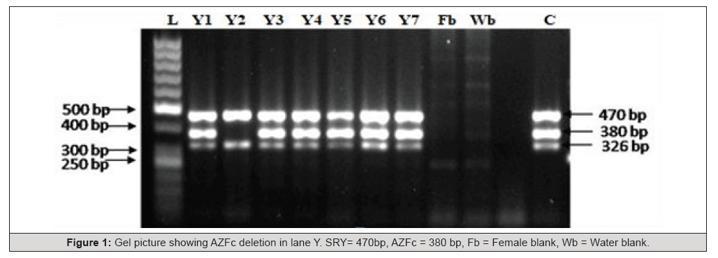
Figure 1:Gel picture showing AZFc deletion in lane Y. SRY= 470bp, AZFc = 380 bp, Fb = Female blank, Wb = Water blank.
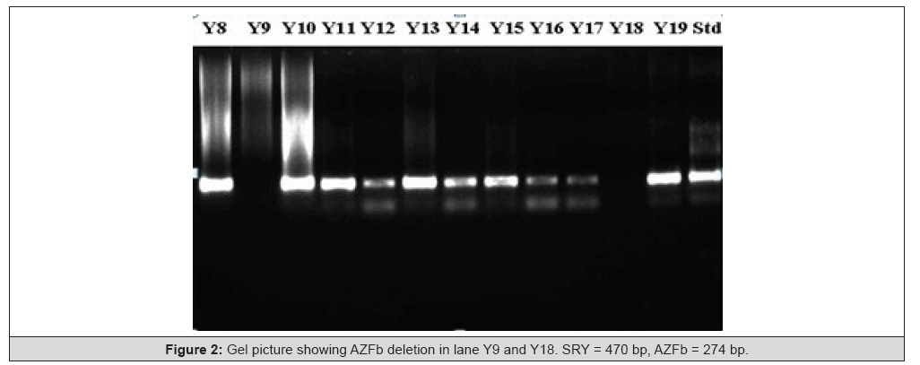
Figure 2:Gel picture showing AZFb deletion in lane Y9 and Y18. SRY = 470 bp, AZFb = 274 bp.
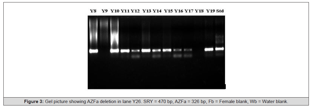
Figure 3:Gel picture showing AZFa deletion in lane Y26. SRY = 470 bp, AZFa = 326 bp, Fb = Female blank, Wb = Water blank.
The serum hormones, viz., FSH, LH, prolactin, testosterone and PSA, levels in azoospermic subjects were 10.08±1.20 mIU/ mL, 4.16±0.82 mIU/mL, 7.86±1.78 ng/mL, 5.15±0.85 ng/mL and 0.90±0.13 ng/mL, respectively. The respective hormone levels in other infertile subjects were 9.23±1.79 mIU/mL, 3.77± 0.89 mIU/ mL, 9.64±1.63 ng/mL, 3.45±0.58 ng/mL and 0.93±0.11 ng/mL, respectively. In azoospermic subjects, serum FSH levels were elevated but not statistically significant, while LH and prolactin levels were significantly increased (p < 0.001). Testosterone levels showed a non-significant decrease compared to other groups, and no notable changes were observed in PSA levels (Table 1). Although hormonal evaluation provides valuable insights, no single hormone can serve as a definitive marker for spermatogenesis.

Table 1:Serum hormone profile of azoospermic subjects and other infertile subjects.
Note*: Data are represented as mean ± SD, *P<0.001, FSH = Follicle stimulating hormone, LH = Luteinizing hormone, PSA = Prostate specific antigen.
FSH remains a key hormone for monitoring infertile men in therapeutic and clinical settings. While it is essential for the initiation of spermatogenesis, an FSH level twice the normal range often indicates germinal epithelial damage. However, both normal and elevated FSH levels can be observed in cases of normal and abnormal spermatogenesis. For instance, elevated FSH may be present in testicular histology such as Sertoli Cell Only Syndrome (SCOS) or hypo spermatogenesis, the latter being characterized by quantitatively reduced spermatogenesis [39].
In our study, azoospermic men with Y chromosome microdeletions exhibited significantly elevated LH and prolactin levels, with a non-significant increase in FSH, consistent with findings reported by Yuen, et al. [35], indicating impaired spermatogenesis. Furthermore, the incidence of chromosomal abnormalities in infertile males ranges from 2% to 8%, increasing to approximately 15% among azoospermic individuals [40-43].
Out of four azoospermic subjects, in one azoospermic subject (Y9) spermatogenesis was arrested at round spermatid stage. The Leydig cells were found normal in size and number. Sloughing of immature germ cells was seen in the lumen (Figure 4). The testicular histology of an azoospermic subject (Y2) displayed Sertoli cells exclusively. Leydig cells were increased in number and exhibited the typical morphology of well-developed cells (Figure 5). The histology of testis of the subject (Y18) showed very few seminiferous tubules and numerous fibroids. Germinal epithelium contained very few germ cells mostly of spermatogonia and spermatocytes (Figure 6). The histology of testis of another subject (Y26) showed seminiferous tubules relatively larger in diameter. The tubules contained highly thick basal lamina and virtually devoid of definite germinal epithelium, Sertoli cells and germ cells were very few. Primary spermatocytes where predominant edema around the germinal cells were evident (Figure 7). Current study shows that one azoospermic subject having microdeletion in AZFc loci shows Sertoli cell only syndrome (SCO) which are confirmatory results with Kamp, et al. [44]. The other azoospermic subject had AZFb loci microdeletion with maturation arrest at spermatid stage is in further agreement with earlier studies [24,45-53].
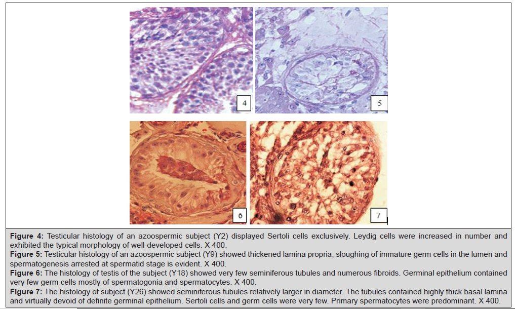
Figure 4:Testicular histology of an azoospermic subject (Y2) displayed Sertoli cells exclusively. Leydig cells were increased in number and
exhibited the typical morphology of well-developed cells. X 400.
Figure 5: Testicular histology of an azoospermic subject (Y9) showed thickened lamina propria, sloughing of immature germ cells in the lumen and
spermatogenesis arrested at spermatid stage is evident. X 400.
Figure 6: The histology of testis of the subject (Y18) showed very few seminiferous tubules and numerous fibroids. Germinal epithelium contained
very few germ cells mostly of spermatogonia and spermatocytes. X 400.
Figure 7: The histology of subject (Y26) showed seminiferous tubules relatively larger in diameter. The tubules contained highly thick basal lamina
and virtually devoid of definite germinal epithelium. Sertoli cells and germ cells were very few. Primary spermatocytes were predominant. X 400.
The idiopathic individuals with severe oligozoospermia or azoospermia have the highest frequency of Yq microdeletion, particularly involving the AZFc region. Testicular histopathological study of the azoospermic subject, showed Y chromosome deletion, revealed complete absence of germ cells and presence of only Sertoli cells. A few reports on Yq microdeletion and infertility in Indian populations are available. A recent study conducted in Kashmir population also supports that gene microdeletion as a major significant cause contributing to the aetiology of infertility among males [54]. In another study on molecular screening for Yq microdeletion with idiopathic oligozoospermia and azoospermia the frequency of microdeletion in northern and eastern India, respectively, was reported to be 9.63% and 8.5% [34,55]. In these studies, deletion in AZFc region was the most common followed by AZFb and AZFa. Ambasudhan, et al. [56] also studied idiopathic cases of male infertility from northern India populations detected a low frequency (5 % only) of Yq microdeletion. In this investigation, AZFc region was also the most frequently deleted region in azoospermic and oligospermic men. In the present study, Yq microdeletion and semen analyse of 30 Indian males seeking for infertility treatment was carried out. However, Yq microdeletion was seen in 2 out of 8 frequencies of microdeletion in azoospermic subject was found to be 6.66% which is nearly similar of the study conducted by Mitra, et al. [57] where they found 5.29% in azoospermic population. The incidence of Y chromosome microdeletion in Hainan infertile men was 7.13%. The occurrence rate of Y chromosome microdeletion was 6.69% in the oligozoospermia group and 7.71% in the azoospermia group [58]. It has been postulated that geographical location and the characteristics of the studied populations are significant factors influencing the observed frequency of Y chromosome microdeletions. These variables can cause deviations from global baseline prevalence rates, resulting in regional variation in microdeletion frequencies [29].
A study by Arumugam, et al. [59] reported a 2% prevalence of Y chromosome microdeletions in the southern Indian population. In contrast, the present study found an overall Yq microdeletion frequency of 13.33% among infertile subjects, which aligns closely with the findings of Najmabadi, et al. [46], who reported an 18% prevalence of Yq microdeletions in cases of idiopathic azoospermia and severe oligozoospermia. Furthermore, Elsaid, et al. [60] conducted a study on Sudanese men, identifying Y chromosome microdeletions localized to the AZFa, AZFb, AZFc and AZFd regions in 58.8% of infertile subjects, with 31.4% azoospermic and 27.5% oligozoospermic. The detection of Yq microdeletion in an infertile man provides a proper diagnosis and allows the clinician to avoid unnecessary and often expensive treatment to improve fertility and has important ethical consequence microdeletions if the patient is a candidate of ART. Microdeletion analysis using PCR helps to determine the frequency and site of gene deletion and thus, testicular phenotype and also contributes to the determination of an accurate prognosis and ultimately to valuable counselling for complies diagnosed with AZF microdeletion. The present study is limited by a relatively small sample size, underscoring the need for further research to refine diagnostic methodologies and develop strategies to accurately detect and prevent DNA damage in spermatozoa utilized in ART.
Conclusion
Our study emphasizes the critical role of comprehensive cytogenetic analysis in all infertile males prior to ART procedures to mitigate the risk of vertical transmission of chromosomal abnormalities. The prevalence of Y chromosome microdeletions among azoospermic patients exhibits variability influenced by ethnic and geographic factors, with deletions predominantly localized within the AZF sub-regions-namely AZFa, AZFb, and AZFc. Advancements in high-throughput genomic and whole-exome sequencing methodologies are anticipated to further delineate the molecular aetiology of spermatogenic failure by identifying novel genetic variants and regulatory elements critical to spermatogenesis. Consequently, rigorous screening of spermatozoa for Y chromosome microdeletions should be integrated as a standard diagnostic criterion before ART application. Additionally, males harbouring Y chromosome microdeletions must be counselled genetically to inform reproductive decision-making and assess the risk of transmission to progeny.
Acknowledgement
TCS, NKL and ASA conceived and designed the study; AS performed the investigations; AS and BK analyzed data and wrote the paper. All authors read and approved the manuscript and all data were generated in-house and that no paper mill was used.
The Head, Department of Zoology, University of Rajasthan, Jaipur (India) provided infrastructural facilities is gratefully acknowledged. The investigation was supported as a major research project (No. 5/10/2/2005-RHN dated 24.2.2006) by the Indian Council of Medical Research, New Delhi (India).
Conflict of Interest
Authors have no conflicts of interest.
References
- Leslie SW, Soon Sutton TL, Khan MAB (2024) Male Infertility. In: StatPearls [Internet]. Treasure Island (FL): StatPearls Publishing, 2025.
- Said TM, Paasch U, Glander H, Agarwal A (2004) Role of caspases in male infertility. Hum Reprod Update 10(1): 39-51.
- Barrera N, Omolaoye TS, Du Plessis SS (2022) A contemporary view on global fertility, infertility, and assisted reproductive techniques. In: Fertility, Pregnancy, and Wellness, Elsevier, pp.93-120. DOI:10.1016/B978-0-12-818309-0.00009-5
- Spano M, Bonde JP, Hjollund HI (2000) Sperm chromatin damage impairs human fertility. Fertil Steril 73(1): 43-50.
- Csenke Z, Török L, Szöllösi J, Scultéty S (1995) Role of endocrine factors in male infertility. Int Urol Nephrol 27: 203-206.
- Charny CW (1940) Testicular biopsy; its value in male sterility. J Amer Med Assoc 115(17): 1429-1432.
- Wong TW, Straus FH, Jones TM, Warner NE (1978) Pathological aspects of the infertile testis. Urol Clin North Am 5(3): 503-530.
- Brannen GE, Roth RR (1979) Testicular abnormalities of the subfertile male. J Urol 122(6): 757-762.
- Nagpal BL, Manjari M, Kapoor K, Dhaliwal US (1993) Testicular biopsy in cases of male infertility: a retrospective study. J Indian Med Assoc 91(7): 171-174.
- Yoshida A, Miura K, Nagao K, Hara H, Ishii N, et al. (1997) Sexual function and clinical features of patients with Klinefelter's syndrome with the chief complaint of male infertility. Int J Androl 20(2): 80-85.
- Rosenlund B, Kvist U, Plöen L, Rozell BL, Sjöblom P, et al. (1998) A comparison between open and percutaneous needle biopsies in men with azoospermia. Hum Reprod 13(5): 1266-1271.
- Jamal A A, Mansoor I (2001) Morphological profile of testicular biopsies associated with infertility. Saudi Med J 22(11): 992-994.
- Franchimont PD, Millet E, Vendrely J, Letawe J, Legros JJ, et al. (1972) Relationship between spermatogenesis and serum gonadotrophin levels in azoospermia and oligospermia. J Clin Endocrinol Metab 34(6): 1003-1008.
- de Kretser DM, Burger HG, Hudson B (1974) The relationship between germinal cells and serum FSH levels in males with infertility. J Clin Endocrinol Metab 38(5): 787-793.
- Yaman O, Ozdiler E, Seckiner I, Gogus O (1999) Significance of serum FSH levels and testicular morphology in infertile males. Int Urol Nephrol 31(4): 519-523.
- Nieschlag E (1993) Care for the infertile male. Clin Endocrinol (Oxf) 38(2): 123-133.
- Ishida H, Isurugi K, Aso Y, Takayasu H, Tamaoki BI (1976) Endocrine studies in Sertoli-cell-only syndrome. J Urol 116(1): 56-58.
- de Kretser DM, Kerr JB, Paulsen CA (1981) Evaluation of the ultrastructural changes in the human Sertoli cell in testicular disorders and the relationship of the changes to the levels of serum FSH. Int J Androl 4(2): 129-144.
- Chandley AC (1979) The chromosomal basis of human infertility. Br Med Bull 35(2): 181-186.
- Faed MJ, Robertson J, Lamont MA, MacIntosh W, Grieve J, et al. (1979) A cytogenetic survey of men being investigated for subfertility. J Reprod Fertil 56(1): 209-216.
- Kjessler B (1974) Chromosomal constitution and male reproductive failure. In: Mancini RE, Martini LC (Editors.), Male Fertility and Sterility. Academic Press, New York, pp. 231-247.
- Goodfellow PN, Lovellbadge R (1993) SRy and sex determination in mammals. Annual Rev Genet 27: 71-92.
- Tiepolo L, Zuffardi O (1976) Localisation of factors controlling spermatogenesis in the nonfluorescent portion of the human Y chromosome long arm. Hum Genet 34(2):119-124.
- Vogt PH, Edelmann A, Kirsch S, Henegariu O, Hirschmann P, et al. (1996) Human Y chromosome azoospermia factors (AZF) mapped to different subregions in Yq11. Hum Mol Genet 5(7): 933-943.
- Vollrath D, Foote S, Hilton A, Brown LG, Beer-Romero P, et al. (1992) The human Y chromosome: A 43-interval map based on naturally occurring deletions. Science 258(5079): 52-59.
- Witherspoon L, Dergham A, Flannigan R (2021) Y-microdeletions: A review of the genetic basis for this common cause of male infertility. Trans Androl Urol 10(3): 1383-1390.
- Krausz C, Rosta V, Swerdloff R, Christina W (2021) Genetics of Male Infertility. In: Pyeritz RE, Korf BR, Grody WW (Editors.), Emery and Rimoin's Principles and Practice of Medical Genetics and Genomics, Perinatal and Reproductive Genetics, (Seventh Edition), Academic Press, London 121-147.
- Foresta C, Moro E, Ferlin A (2001): Y chromosome microdeletions and alterations of spermatogenesis. Endocr Rev 22(2): 226-239.
- Krausz C, McElreavey K (2001): Y chromosome microdeletions in ‘fertile’ males. Hum Reprod 16(6): 1306-1307.
- World Health Organization (1999) Laboratory Manual for the Examination of the Human Semen and Sperm Cervical Mucus Interaction. Cambridge University Press, Cambridge.
- Laird PW, Zijderveld A, Linders K, Rudnicki MA, Jaenisch R, et al. (1991) Simplified mammalian DNA isolation procedure. Nucleic Acids Res 19(15): 4293.
- Sambrook J, Russell DW (2001) Molecular Cloning; A Laboratory Manual, (III Edition), Vol. 1, Cold Spring Harbor Laboratory Press, New York, pp. 6.8-6.10.
- Simoni M, Bakker E, Eurlings MC, Matthijs G, Moro E, et al. (1999) Laboratory guidelines for molecular diagnosis of Y-chromosomal microdeletions. Int J Androl 22(5): 292-299.
- Dada R, Gupta NP and Kucheria K (2003) Molecular screening for Yq microdeletion in men with idiopathic oligozoospermia and azoospermia. J Biosci 28(2): 163-168.
- Yuen W, Golin AP, Flannigan R, Schlegel PN (2021) Histology and sperm retrieval among men with Y chromosome microdeletions. Transl Androl Urol 10(3): 1442-1456.
- Sultana S, Nallari P, Ananthapur V (2020) Recurrent pregnancy loss (RPL): An overview. J Women’s Health Dev 3(3): 302-315.
- Rabinowitz MJ, Huffman PJ, Haney NM, Kohn TP (2021) Y‐chromosome microdeletions: A review of prevalence, screening, and clinical considerations. Appl Clin Genet 14: 51-59.
- Pazoki N, Salehi M, Angaji SA, Abdollahpour Alitappeh M (2024) Association of Y chromosome AZF region microdeletions with recurrent miscarriage in Iranian couples: A case-control study. Mol Genet Genomic Med 12(2): e2392.
- Abid S, Maitra A, Meherji P, Patel Z, Kadam S, et al. (2008) Clinical and laboratory evaluation of idiopathic male infertility in a secondary referral center in India. J Clin Lab Anal 22(1): 29-38.
- Foresta C, Bettella A, Merico M, Garolla A, Ferlin A, et l. (2002) Use of recombinant human follicle-stimulating hormone in the treatment of male factor infertility. Fertil Steril 77(2): 238-244.
- Huynh T, Mollard R, Trounson A (2002) Selected genetic factors associated with male infertility. Hum Reprod Update 8(2): 183-198.
- Vicdan A, Vicdan K, Günalp S, Kence A, Akarsu C, et al. (2004) Genetic aspects of human male infertility: the frequency of chromosomal abnormalities and Y chromosome microdeletions in severe male factor infertility. Eur J Obstet Gynecol Reprod Biol 117(1): 49-54.
- Mau-Holzmann UA (2005) Somatic chromosomal abnormalities in infertile men and women. Cytogenet Genome Res 111(3-4): 117-336.
- Kamp C, Huellen K, Fernandes S, Sousa, Schlegel MPN, et al. (2001) High deletion frequency of the complete AZFa sequence in men with Sertoli-cell-only syndrome. Mol Hum Reprod 7(10): 987-994.
- Reijo R, Lee TY, Salo P, Alagappan R, Brown LG, et al. (1995) Diverse spermatogenic defects in humans caused by Y chromosome deletions encompassing a novel RNA-binding protein gene. Nat Genet 10(4): 383-393.
- Najmabadi H, Huang V, Yen P, Subbarao MN, Bhasin D, et al. (1996) Substantial prevalence of microdeletions of the Y-chromosome in infertile men with idiopathic azoospermia and oligozoospermia detected using a sequence-tagged site-based mapping strategy. J Clin Endocrinol Metab 81(4): 1347-1352.
- Qureshi SJ, Ross AR, Ma K, Cooke HJ, Intyre MA, et al. (1996) Polymerase chain reaction screening for Y chromosome microdeletions: a first step towards the diagnosis of genetically-determined spermatogenic failure in men. Mol Hum Reprod 2(10): 775-779.
- Stuppia L, Mastroprimiano G, Calabrese G, Peila R, Tenaglia R and Palka G (1996): Microdeletions in interval 6 of the Y chromosome detected by STS-PCR in 6 of 33 patients with idiopathic oligo- or azoospermia. Cytogenet Cell Genet 72(2-3): 155-158.
- Grardi SK, Meilnik, Schegel PN (1997) Submicroscopic deletions in the Y chromosome of infertile men. Hum Reprod 12(8): 1635-1641.
- Grimaldi P, Scarponi C, Rossi P, Rocchietti March M, Fabbri A, et al. (1998) Analysis of Yq microdeletions in infertile males by PCR and DNA hybridization techniques. Mol Hum Reprod 4(12): 1116-1121.
- Lee JH, Lee DR, Yoon SJ, Chai YG, Roh SI, et al. (1998) Expression of DAZ (deleted in azoospermia), DAZL1 (DAZ-like) and protamine-2 in testis and its application for diagnosis of spermatogenesis in non-obstructive azoospermia. Mol Hum Reprod 4(9): 827-834.
- Kleiman SE, Yogev L, Gamzu R, Hauser R, Botchan A, et al. (1999) Three-generation evaluation of Y-chromosome microdeletion. J Androl 20(3): 394-398.
- Krausz C, Quintana-Murci L, Barbaux S, Siffroi JP, Rouba H, et al. (1999) A high frequency of Y chromosome deletions in males with nonidiopathic infertility. J Clin Endocrinol Metab 84(10): 3606-3612.
- Ahmad F, Zargar MH, Lateef M, Hussain A, Malla TM, et al. (2024) Azoospermia factor gene microdeletions in infertile men with non-obstructive azoospermia and normal karyotype: First case-control study from Kashmir. Gene Reports 37(1):102064.
- Thangaraj K, Subramanian S, Reddy AG, Singh L (2003) Unique case of deletion and duplication in the long arm of the Y chromosome in an individual with ambiguous genitalia. Am J Med Genet 116A(2): 205-207.
- Ambasudhan R, Singh K, Agarwal JK, Singh SK, Khanna A, et al. (2003) Idiopathic cases of male infertility from a region in India show low incidence of Y-chromosome microdeletion. J Biosci 28(5): 605-612.
- Mitra A, Dada R, Kumar R, Gupta NP, Kucheria K and Gupta SK (2008) Screening for Y-chromosome microdeletions in infertile Indian males: Utility of simplified multiplex PCR. Indian J Med Res 127(2): 124-132.
- He Q, Zhang Y, Song M, Zhou Y, Lin D, et al. (2024) Detection of AZF microdeletions and analysis of reproductive hormonal profiles in Hainan men undergoing assisted reproductive technology. BMC Urol 24(1): 123.
- Arumugam M, Shetty DP, Kadandale JS, Kumari SN (2021) Y chromosome microdeletion and cytogenetic findings in male infertility: Across-sectional descriptive study. Int J Reprod BioMed 19(2): 147-156.
- Elsaid HOA, Gadkareim T, Abobakr T, Mubarak E, Abdelrhem MA, et al. (2021) Detection of AZF microdeletions and reproductive hormonal profile analysis of infertile Sudanese men pursuing assisted reproductive approaches. BMC Urology 21(1): 69 (1-9).




 We use cookies to ensure you get the best experience on our website.