
Case Report 
 Creative Commons, CC-BY
A Case of Pericardial Effusion in A Patient with Pace- Maker and Covid-19 Pneumonia at MSCT
*Corresponding author:Francesco Messina, MD, Unit of Radiology - Riuniti Hospital, Great Metropolitan Hospital (G.O.M.) “Bianchi–Melacrino– Morelli”, Via Giuseppe Melacrino n.21, 89124 Reggio Calabria – Italy.
Received:August 20, 2025; Published:August 25, 2025
DOI: 10.34297/AJBSR.2025.28.003666
Abstract
Pericardial effusion is a well-known complication of several viral infections, such as influenza virus, coxsackievirus and echovirus. But there is also a correlation between Covid-19 coronavirus and related pericardial effusion. Here we describe the case of a 67-years-old man, that had precedently an ablation for atrial fibrillation (in other Hospital), and Pace-Maker dependence just for this arrhythmia, that also had a bilateral Covid-19 pneumonia complicated by pericardial effusion. An unenhanced Computed Tomography was urgently made, to treat the patient and for monitoring his clinical conditions.
Keywords:Pericardial Effusion, Pace-Maker, Covid-19 Pneumonia, MSCT
Introduction
In these years the world had faced the terrible Covid-19 coronavirus pandemic emergency, with a rapidly expanding pandemic of lower respiratory tract infection. The heterogeneity of the disease course poses a challenge to healthcare providers and optimal management of the patients. The use of CT imaging in the diagnosis and follow-up had rapidly grown, and radiological patterns along the disease course are increasingly understood [1], and had established itself with numerous evaluation patterns of the Covid 19 disease, which can therefore be characterized as a multi-organ disease. CT is still considered the first-line imaging tool regarding SARS-CoV-2 infection [2]. This case highlights the utility of CT in detecting pericardial effusion in a Covid-19 patient with preexisting Pace-Maker device.
Case Presentation
In a 67-years-old man suffering from atrio-ventricular block and with a Pace-Maker already implanted in the left thorax, nasopharyngeal sampling was positive for SARS-CoV-2. He had persistent chest pain (retrosternal, intensified when coughing and lay ing down), shortness of breath, and fever (39°C) since about four days, without clinical improvements from taking paracetamol.
He arrived to the Emergency Department of our Hospital, where he had:
a. negative Ecg for ischemic alterations;
b. auscultation: vescicular murmur was reduced in bi-basal
lungs;
c. sO2 of 88%;
d. Laboratory: leukopenia and increased values of C-reactive
protein.
He had urgently made a chest Computed Tomography (High Resolution Computed Tomography, HRCT), without contrast, that showed (Figure 1, a-b) the presence of typical “Ground-Glass” (GGO) bi-basal areas in both lungs, with bilateral consolidations areas, and also the evidence of bilateral pulmonary interstitial thickenings. All these CT signs were suggestive for bilateral Covid19 pneumonia (Figure 1).
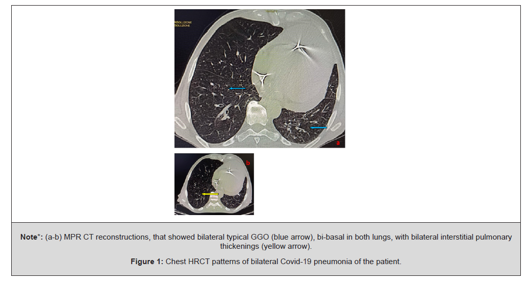
Note*: (a-b) MPR CT reconstructions, that showed bilateral typical GGO (blue arrow), bi-basal in both lungs, with bilateral interstitial pulmonary thickenings (yellow arrow).
Figure 1:Chest HRCT patterns of bilateral Covid-19 pneumonia of the patient.
CT also showed (Figure 2, a-b) the presence of a very large pericardial effusion (which is arranged around the heart, with maximum thickness of about 4cm), especially identifiable in the antero- lateral and apical locations, with an enlargement of the heart. An other relief was the obliteration and thickening of the mediastinal adipose plane anteriorly. HRCT also showed very well the important finding, as the presence of cardiac Pace-Maker (Figure 2).
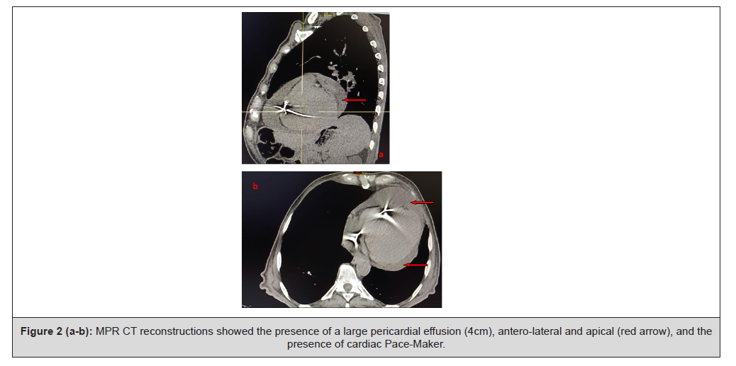
Figure 2 (a-b):MPR CT reconstructions showed the presence of a large pericardial effusion (4cm), antero-lateral and apical (red arrow), and the presence of cardiac Pace-Maker.
The patient was immediately hospitalized, and, after about two weeks of hospitalization, he had returned at home. Now the patient will subsequently perform a clinical-radiological and laboratory follow-up.
On the Space Fp of Proton’s Wave Functions
Critical patients with Covid-19 pneumonia showed higher incidences of pericardial effusion, pleural effusion and lymphnodes enlargements than ordinary patients [3]. The pathogenesis of Covid-19 pericardial effusion is yet unresolved; two predominant mechanisms could be relevant [4]. First, the heart affinity of the virus could be explained by SARS-CoV-2 S protein direct binding to human angiotensin-converting enzyme 2 present in the human heart, which allows for a cellular infection. Indirectly, pericardial effusion could follow a viral replication and dissemination in the blood, from day 7 up to 1 month after symptoms beginning. This could lead to a cytokine storm syndrome and a direct myopericardial lesion by inflammatory cell infiltration, similarly to Covid-19 direct pulmonary lesions [5-6]. Current guidelines advocate the use of unenhanced chest CT for the diagnosis, severity assessment and monitoring of Covid-19 disease [7]. Computed Tomography is important not only in the early stages of Covid-19 diagnosis, but also in the evaluation of complications (such as pleural-pericardial effusion) and other concomitant pathologies during the subsequent course of the disease, orienting toward the best clinical management/ outcome for the patients [8]. And Computed Tomography had an important role in the diagnosis and severity evaluation of the disease, because it investigates very well the dynamic CT changes in different stages of Covid-19 pneumonia, and is very important in the follow-up of the patients [9]. Moreover, pericardial effusion and tamponade as a complication of epicardial lead placement had been rarely described [10,11]. Pericardial effusion is provoked by a variety of infectious and non-infectious processes. The primary aim in treating patients with symptomatic pericardial effusion is relief of the symptoms.
Conclusion
This case underscores the importance of chest CT in identifying pericardial effusion in Covid-19 patients, particularly those with pre-existing cardiac devices, where symptoms may be attributed to multiple causes. What made the diagnosis of pericardial effusion unclear in this patient was the competing, already established diagnoses that warranted him Hospital admission and epicardial device placement in the first place: Covid-19 pneumonia. Moreover, pericardial effusion is a very important condition must be promptly identified, treated and monitored over time. Cross-sectional imaging techniques such as Computed Tomography, however, can display the detailed anatomic features of the entire pericardium more clearly than other methodics, and CT has been found to be an excellent tool for detecting pericardial effusion and its clinical course.
Conflicts of Interest
The authors certify that there is no conflict of interest with any financial organization regarding the material discussed in the manuscript.
Patient Consent Statement Mandatory
The patient confirmed the consense for publication of our case report.
References
- Tao Ai, Zhenlu Yang, Hongyan Hou, Chenao Zhan, Chong Chen, et al. (2020) Correlation of chest CT and RT-PCR testing in Coronavirus Disease 2019 (COVID-19) in China: a report of 1014 cases. Radiology. 296(2): E32-E40.
- ACR recommendations for the use of chest radiography and computed tomography for suspected Covid-19 infection. https://www.acr.org/Advocacy-and-Economics/ACR-Position-Statements/Recommendations-for-ChestRadiographyand-CT-for-Suspected-Covid-19-Infection.
- Dong N, Cai J, Zhou Y, Liu J, Li F (2020) End-stage heart failure with COVID-19: strong evidence of myocardial injury by 2019-nCoV. JACC Heart Fail 8(6): 515–517.
- Richa Purohit, Arjun Kanwal, Anil Pandit, Bhavin M Patel, Glenn Robert Meininger (2020) Acute myopericarditis with pericardial effusion and cardiac tamponade in a patient with Covid-19. Am J Case Rep 21: e925554.
- Asif T, Kassab K, Iskander F, Alyousef T (2020) Acute pericarditis and cardiac tamponade in a patient with Covid-19: a therapeutic challenge. Eur J Case Rep Intern Med 7(6): 001701.
- Inciardi RM, Lupi L, Zaccone G, Italia L, Raffo M, Tomasoni D, et al. (2020) Cardiac involvement in a patient with coronavirus disease 2019 (COVID-19). JAMA Cardiol 5(7): 819-824.
- Kunhua Li, Jiong Wu, Faqi Wu, Dajing Guo, Linli Chen, et al. (2020) The clinical and chest CT features associated with severe and critical Covid-19 pneumonia. Invest Radiol 55(6): 327-331.
- Messina F, Turano L, Tebala C, Calabrese G, Arcadi N (2021) Pericardial and pleural effusion in an elderly woman with Covid-19 pneumonia: CT findings. Radiol Case Rep 16(6): 1335-1338.
- Messina F, Tebala C, Turano L, Calabrese G, Arcadi N (2020) The spider web sign in Covid-19 pneumonia: An interesting case studied to resolution with Computed Tomography. Radiol Case Rep 16(3): 673-677.
- Peters RW, Scheinman MM, Raskin S, Thomas AN (1980) Unusual complications of epicardial pacemakers: recurrent pericarditis, cardiac tamponade and pericardial constriction. Am J Cardiol 45(5): 1088-1094.
- Schwartz DJ, Thanavaro S, Kleiger RE, Krone RJ, Connors JP, et al. (1979) Epicardial Pace-Maker complicated by cardiac tamponade and constrictive pericarditis. Chest 76(2): 226-227.




 We use cookies to ensure you get the best experience on our website.