
Opinion 
 Creative Commons, CC-BY
3D Guided Screw Placement in the Spine and Pelvis – Who Bears Responsibility? – A User’s Perspective
*Corresponding author:Axel Jubel, Department of Trauma- and Reconstructive Surgery, Eduardus Hospital Cologne/Germany, Custodis-Str. 3-17, 50679 Cologne/Germany.
Received:August 18, 2025; Published:August 21, 2025
DOI: 10.34297/AJBSR.2025.28.003659
Abstract
The use of three-dimensional (3D)- guided navigation and optical tracking in spine and pelvic surgery has significantly evolved over the past two decades. This technology enhances implant placement accuracy, improves surgical outcomes, and reduces intraoperative complications such as neurological and vascular injuries. Additionally, it enables minimally invasive techniques, lowers radiation exposure for the surgical team, and improves implant anchorage, particularly in osteoporotic bone. Despite these advantages, the surgeon remains fully responsible for the procedure’s success. Accurate referencing, continuous validation of virtual versus real anatomy, and the ability to proceed without navigation in case of system failure are critical. This paper provides a user-centered perspective on the practical applications, benefits, limitations, and medico-legal implications of 3D Guided intraoperative navigation.
Keywords:3D Navigation, Artificial Intelligence in Surgery, Pedicle Screw Placement, Minimally Invasive Spine Surgery, Radiation Exposure Reduction, Responsibility in Surgery
Abbreviations:AI: Artificial Intelligence
Introduction
For more than 20 years, trauma surgery has benefited from artificial intelligence (AI) in the form of 3D-guided screw placement in the spine and pelvis. While the early stages were laborious and led to significantly longer operative times compared to conventional techniques, advancements in hardware and software have made possible a user-friendly surgical aid now appreciated by many surgeons.
To perform 3D-guided navigation, a powerful computer system with appropriate AI software, connection to an optical tracking system, and a 3D-capable C-arm fluoroscope is required.
A navigation-guided procedure essentially involves three steps: first, the placement of a reference clamp; second, acquisition of the 3D dataset; and finally, virtual placement of the implants within the 3D image. To allow the optical system to detect the reference clamp and registered instruments, these are equipped with small reflective spheres. The primary applications of 3D-guided intraop erative navigation in trauma surgery are in the spine and pelvis. In these anatomically complex regions, conventional two-dimensional imaging is associated with a high rate of error, particularly in the minimally invasive placement of stabilizing implants [1-3].
For example, in the case of stabilizing a vertebral body fracture, the surgeon must place a screw with a diameter of 4.5 to 7.5 mm into each pedicle of the adjacent vertebrae. Pedicles themselves often measure only 5–8 mm in diameter, leaving minimal margin for error. The screw must pass through the center of the pedicle to ensure sufficient anchorage. Perforation of the medial pedicle wall risks spinal cord injury, while exiting cranially or caudally may compromise nerve roots. Anterior cortical breach may result in life-threatening vascular injury.
Patients benefit primarily from intraoperative computer navigation, which provides increased safety. Numerous comparative studies have shown that 3D computer-navigated screw placement enables significantly more precise implant positioning than conventional methods [1-3]. As a result, complications due to screw misplacement—such as spinal cord, nerve, or vascular injuries— occur far less frequently [4]. Three-dimensional visualization of anatomical structures allows screws to be placed through very small incisions, making extensive exposure of the surgical site unnecessary. It is well established that minimally invasive procedures result in less postoperative pain, shorter recovery times, and lower rates of wound healing disorders and infections. Another advantage for the patient is that accurate placement of implants in dense bone provides secure anchorage, reducing the likelihood of postoperative loosening [4].
Benefits for the Surgeon and OR Team
In conventional procedures, the surgeon relies heavily on repeated intraoperative fluoroscopic imaging to guide pedicle or pelvic screw placement. During imaging, the entire surgical team is exposed to radiation, often accumulating fluoroscopy times in the double-digit minute range per patient. Teams performing two to three spinal procedures daily are subjected to high cumulative annual radiation exposure.
In 3D-guided computer navigation, the required 3D images are acquired preoperatively, immediately after patient positioning and before the skin incision. During this time, the surgical team exits the room. Intraoperative fluoroscopy is only used briefly for verification, resulting in significantly reduced radiation exposure per procedure and cumulatively [1-3].
Surgeons also benefit from superior visualization of their operative actions. They can see the effect of any change in instrument position on the virtual 3D anatomy before making the actual move. Even minimal adjustments can result in a significantly better final implant position. This skill must be learned, but with frequent repetition, the learning curve is steep. Surgeons often show rapid improvement in hand-eye coordination [5].
In osteoporotic patients, conventional imaging often fails due to the low-density bone structures of the spine and pelvis and interference from surrounding soft tissue and air. Poor contrast makes anatomical boundaries difficult to discern. However, 3D imaging can clearly visualize bone structures without overlap, even in the presence of osteoporotic changes. Navigation thus provides the surgeon with clarity and speed, as the anatomy remains consistently identifiable.
Potential Pitfalls and Responsibility
For 3D-guided navigation to function correctly, a reference clamp must be securely attached to a spinous process. From this reference, the AI continuously calculates the position of the instruments relative to the anatomical structures acquired in the 3D dataset. Any movement of the reference clamp after image acquisition alters the relationship between virtual and real anatomy. Therefore, the surgeon must regularly verify that the virtual anatomy still matches the real anatomy [1]. This is typically done by placing a registered instrument on a known anatomical landmark. Since the system displays both the surface and intrabony position of the instrument, alignment of virtual and real anatomy should also be checked through brief fluoroscopic verification.
If discrepancies are identified, corrections may be necessary. If the reference clamp has been displaced, a new 3D dataset may need to be acquired before the procedure can continue. If a malfunction of the navigation system is detected, the procedure may have to be completed without navigation. Thus, the surgeon must be capable of performing the surgery without navigational assistance if required.
Preoperatively, patients must be informed not only about the planned use of 3D AI-guided navigation but also about the possibility that the procedure may need to be completed without the assistance of AI.
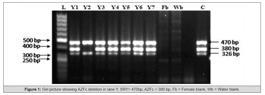
Figure 1:Gel picture showing AZFc deletion in lane Y. SRY= 470bp, AZFc = 380 bp, Fb = Female blank, Wb = Water blank.

Table 1:Serum hormone profile of azoospermic subjects and other infertile subjects.
Note*: Data are represented as mean ± SD, *P<0.001, FSH = Follicle stimulating hormone, LH = Luteinizing hormone, PSA = Prostate specific antigen.
Conclusion
From a user’s perspective, responsibility for the safe execution of 3D-guided, AI-assisted navigation lies entirely with the surgeon— from secure placement of the reference clamp to 3D image acquisition, to final implant positioning. The surgeon must be able to recognize inconsistencies or system malfunctions and be prepared to continue the procedure without navigation if necessary.
Conflict of Interest
None.
References
- Richter PH, Gebhard F (2023) Application of navigation in the fractured spine. Oper Orthop Traumatol 35(1): 29-36.
- Shuman WH, Valliani AA, Chapman EK, Martini ML, Neifert SN, et al. (2022) Intraoperative Navigation in Spine Surgery: Effects on Complications and Reoperations. World Neurosurg 160: e404-e411.
- Tajsic T, Patel K, Farmer R, Mannion RJ, Trivedi RA (2018) Spinal navigation for minimally invasive thoracic and lumbosacral spine fixation: implications for radiation exposure, operative time, and accuracy of pedicle screw placement. Eur Spine J 27(8): 1918-1924.
- Innocenzi G, Bistazzoni S, D'Ercole M, Cardarelli G, Ricciardi F (2017) Does Navigation Improve Pedicle Screw Placement Accuracy? Comparison Between Navigated and Non-navigated Percutaneous and Open Fixations. Acta Neurochir Suppl 124: 289-295.
- Guha D, Moghaddamjou A, Jiwani ZH, Alotaibi NM, Fehlings MG, et al. (2019) Utilization of Spinal Intra-operative Three-dimensional Navigation by Canadian Surgeons and Trainees: A Population-based Time Trend Study. Can J Neurol Sci 46(1): 87-95.




 We use cookies to ensure you get the best experience on our website.